From the whole head to a single synapse: real anatomy, real physics and real data at every scale, rendered at cinematic quality. Free and open, for research and education.
The app needs a WebGPU browser: desktop Chrome or Edge, or recent Safari.
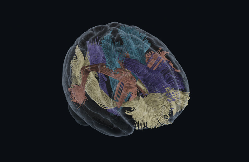
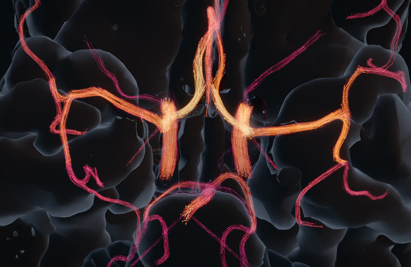
MethodsHCP1065 population tractography (Yeh 2022), 503,085 streamlines resident and drawn as lit tubes with fibre-scale occlusion under a distance LOD, on the MNI152 2009c cortex · wgpu engine, native 2940×1912 capture, M1 Max
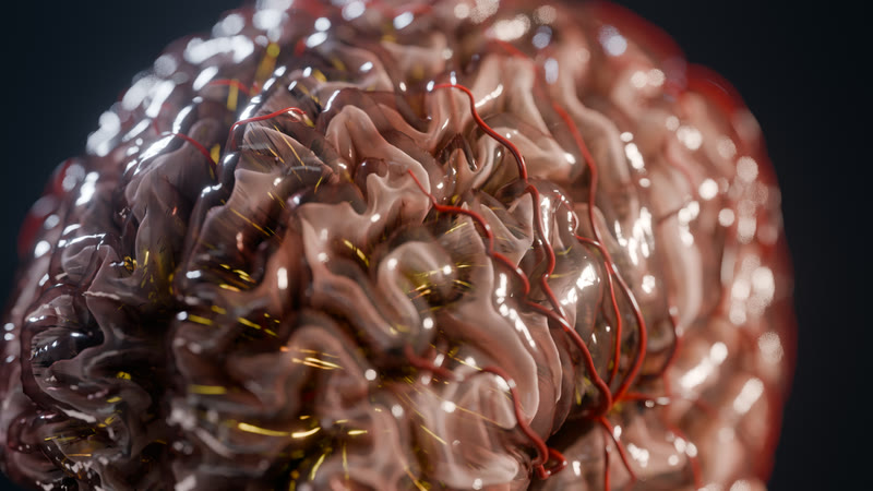
MethodsThe Living Brain, hero v1 · BigBrain's own cortex (Amunts et al. 2013) turning translucent over the HCP1065 fibre bundles, arteries pulsing with the 1D model · Blender 5.2 Cycles, 2560×1440
The 90-second reel, rough cut v2. Six sequences in order, from the whole head to one receptor, cut from the stills and test clips that exist today; the seizure, stroke and synapse stills are the finished 1440p frames. Every segment swaps for its final as it lands. Tap to watch.
MethodsCut from hero-v1, the sequences' 1440p stills and 720p clips with caption cards from their render sidecars, 0.5 s dissolves · 21 segments, 1920×1080, 24 fps, 89.6 s
Scene 1 · Powers of Ten
One continuous zoom, real data at every scale.
A whole cortex, then BigBrain's stained sections at 100 µm, then H01's electron-microscopy neurons: one push, no cuts, every pixel from a measured dataset.
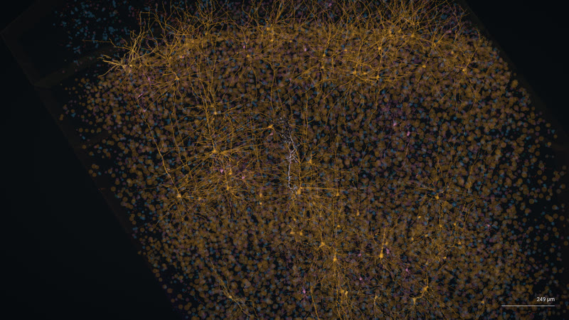
From the cortex to one neuron in an 18-second push. BigBrain's histology dissolves into H01's column of 7,698 reconstructed cells, 2,741 of them neurons, and settles on a single proofread cell.
Methodswgpu engine, native Metal capture at 1080p · BigBrain 2015 (Amunts et al. 2013) at 100 µm, ray-marched bricks · H01 human temporal cortex EM, pack h01-patch-v0 0.2.1: 7,698 skeletons, 5.2 M synapse rows
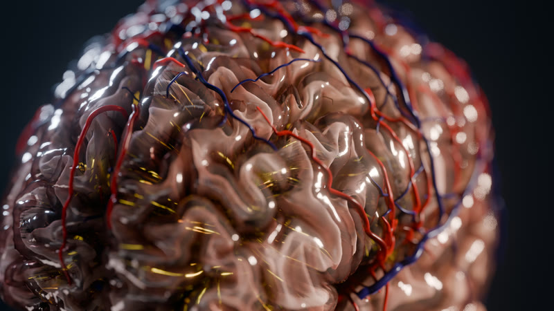
The Living Brain, frame 220. BigBrain's cortex turning to glass over its fibre bundles and arteries, lit like tissue.
MethodsBigBrain pial surface (163,842 vertices a hemisphere) · 39,966 HCP1065 streamlines from 77 bundles · Z-Anatomy arteries (214 centrelines) pulsing with the CoW 1D v0.1 model · Blender 5.2 Cycles, 1024 samples, 2560×1440
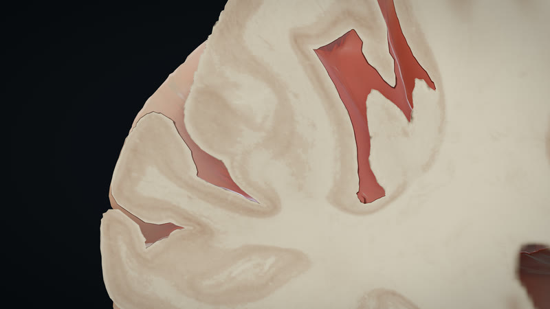
BigBrain at 100 µm, cut open. Real histology as a bricked volume, ray-marched in the engine with a moving section.
MethodsBigBrain 2015, 100 µm volume as a brick pyramid · wgpu engine, native Metal, 1080p
The stain, up close. Cell-dense cortex against pale white matter in BigBrain's own sections, captured at 4K.
Every bundle in the atlas. 503,085 streamlines resident in four anatomical colour families, drawn as lit tubes with fibre-scale occlusion, depth fog and a distance LOD.
MethodsHCP1065 (Yeh 2022), 91 bundles simplified to 15.5 M points at 0.1 mm · wgpu engine, 2940×1912
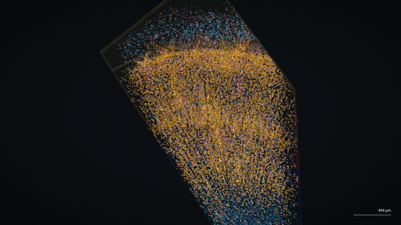
Inside H01's column. Thousands of reconstructed human neurons from a cubic millimetre of temporal cortex, imaged at 4 × 4 × 33 nm.
One proofread cell. The push ends on a single H01 neuron with its arbor, among the dim somata of its neighbours. Scale bar 249 µm.
MethodsH01 proofread morphology · BigBrain cortex and histology behind it · wgpu engine
Scenes 2 and 3 · Blood flow and stroke
Pulsatile flow through one real person's arteries.
A 1D network of 88 arteries everywhere and 3D CFD in the hero vessels, on IXI165's own angiogram. Then a clot lodges, the collaterals take over, and the clock starts on the tissue.
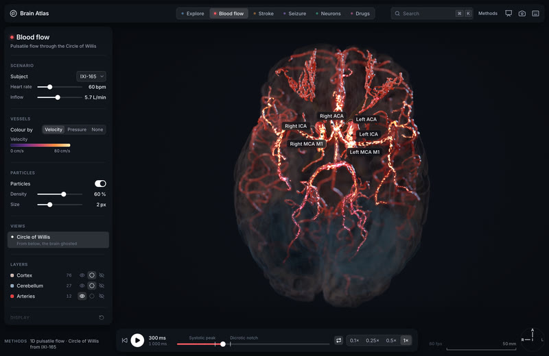
20,000 tracers ride the heartbeat. Drawn from the CFD's 100,000, through IXI165's Circle of Willis for one simulated cycle, looped three times: fast in the carotids, slow and swirling at the apices, inside a membrane that glows with wall shear stress.
Methods3D lattice-Boltzmann CFD at 0.1 mm on IXI165's TOF-MRA lumen (242,682-vertex wall), inlets from the 1D v4 model, cycle periodic to 0.3 % · Blender 5.2 Cycles, 1080p at 4.17× slower than life
794mL/mintotal cerebral flow in the model; 4D flow MRI puts the reference at 717 ± 123
88arteries in the 1D network, from this person's TOF-MRA
0.1mmCFD lattice spacing: 24 cells across the carotid radius
30% vs 13%of the lenticulostriate bed lost to proximal versus distal M1 clots in 72 real patients (p = 0.0026)
The same network, live in the engine. Particles coloured by velocity, pulsing with the 1D solution, in the wgpu viewport.
MethodsopenBF 1D network v4 on IXI165's vessels, checked against PC-MRI · wgpu engine, 1080p orbit
A clot in the left middle cerebral artery: before and after. Distal M1 occlusion. The anterior communicating artery's flow rises from 57 to 86 mL/min as the circle compensates.
Methodsixi165-1d-v4-occl-l-m1 against the v4 baseline · wgpu engine, split capture stacked for the phone
Left carotid blocked: the circle reverses. The left A1 turns around (+134 to −111 mL/min) and 216 mL/min crosses the anterior communicating artery from right to left.
Methodsixi165-1d-v4-occl-l-ica, 15 s · Blender 5.2 Cycles
A basilar clot, and one tiny artery decides. With the traced posterior communicating artery the tissue is spared; with the main anatomy the untreated infarct reaches 153 mL by 24 hours.
Methodsixi165-1d-v4-occl-ba with the PCoA bracket · stroke-v4 tissue fate on IXI165's territories · Blender 5.2 Cycles
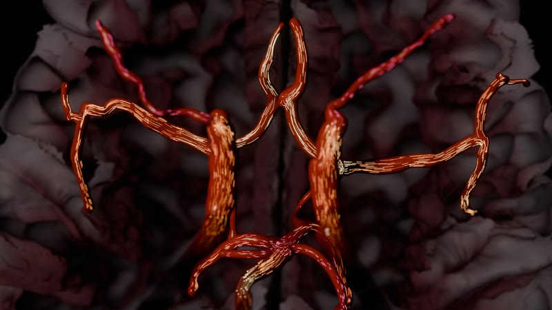
The circle, pulsing, under film light. Particles ride the simulated velocities through this person's arteries while the walls swell with each beat.
MethodsIXI165 TOF-MRA centrelines with TopCoW labels · run ixi165-1d-v4-baseline (openBF), Lagrangian particles on a power-law profile · Blender 5.2 Cycles, 128 samples, shot 2a frames 1–72
The Circle of Willis from this person's own angiogram. Centrelines and radii from the TOF-MRA where the tree overlaps the network, with the flow's particles inside.
MethodsIXI165 (IXI, 3 T TOF-MRA), TopCoW labels · run ixi165-1d-v4-baseline · Blender 5.2 Cycles, 1024 samples
Peak systole, in the engine. The hero camera on the v4 flow at the top of the pulse.
Methodswgpu engine, flow v4 still, 2940×1912
Arteries and veins on the living cortex. Red pial arteries and blue veins on BigBrain's surface, the fibre bundles glowing through the tissue, the arterial tree swollen by the simulated pressure of one heartbeat.
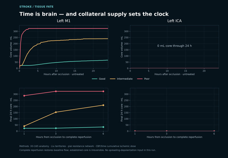
Time is brain, in millilitres. Proximal M1 clot with intermediate collaterals: reperfusion at 1 h prevents 199 mL of the 24-hour infarct, at 3 h 87 mL, at 6 h 29 mL.
MethodsTissue-fate model on IXI165's territories over the 1D v4 occlusion; untreated 24 h core 240 mL
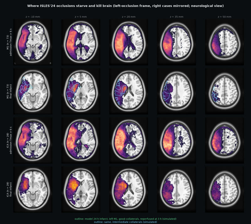
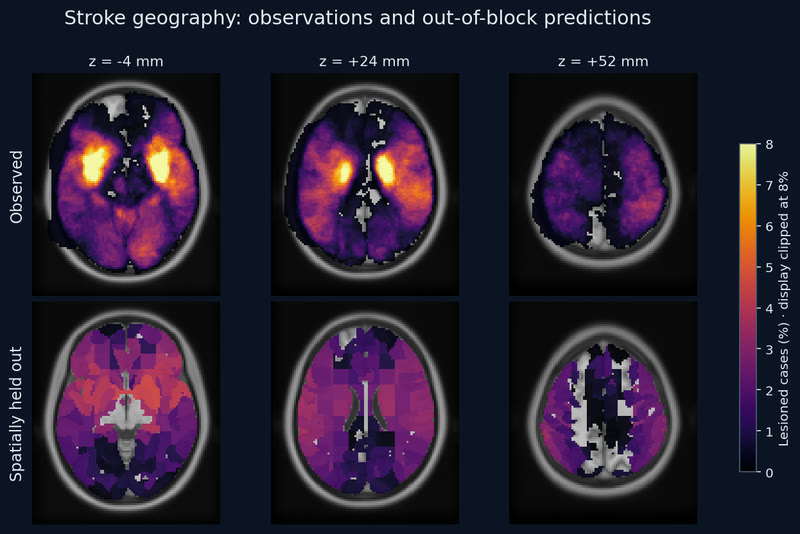
Where real strokes starve and kill brain. 149 ISLES'24 patients: at admission the Tmax > 6 s deficit fills the MCA territory; after thrombectomy the final infarct concentrates in the deep lenticulostriate bed, hit in 47 % of M1 and 57 % of ICA patients at the peak voxel.
MethodsISLES'24 training set, 149 cases registered to MNI (brain-mask Dice median 0.977); outlines are the model's 24 h infarct for a left M1 clot
A spreading depolarization crosses one person's cortex. After the left M1 occlusion, a simulated depolarization wave spreads for 30 minutes until it covers a quarter of IXI165's left cortex. The engine draws it live on that person's own folds, under their own arteries.
MethodsFitzHugh-Nagumo reaction-diffusion on fsaverage, seeded by the v4 left-M1 penumbra (spreading-depolarization-ixi165-m1-v4-8563700d-t0-v1), mapped onto ixi-165-cortex-hd 0.1.0 (IXI165's FastSurfer pial surface) through its map-fsaverage; wgpu engine, native 1080p, 150× model time. · simulated
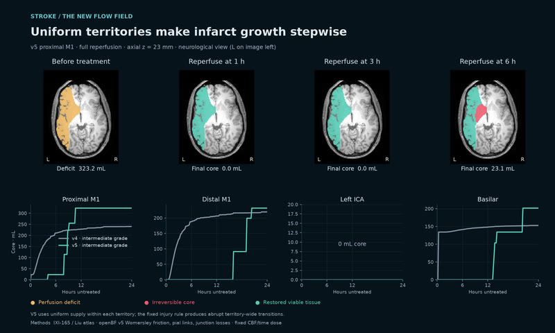
Uniform flow makes infarct growth stepwise. With the uniform v5 tissue rule, proximal-M1 core is 0, 0 and 23.1 mL after reperfusion at 1, 3 and 6 h.
MethodsIXI-165 geometry and Liu territories; openBF v5 with Womersley friction, literature pial links and total-pressure junctions, coupled to the fixed relative-CBF/time injury rule. V4 intermediate grade is compared with v5, whose three grades are identical. Neurological view: left is image left. · simulated
Scene 4 · Seizure
A focal onset recruits the cortex.
A seizure starts in the left mesial temporal lobe and spreads, with the scalp EEG and the depth electrodes a clinician would see. Real patients sit beside the model.
The seizure spreads across the cortex. Each parcel pulses with its own simulated discharges as the network recruits, played at 2.2× model time.
MethodsEpileptor on HCP-derived connectivity, fsaverage cortex, MNE BEM scalp EEG · wgpu engine with the bloom pass, 1080p
The same seizure under film light. BigBrain's cortex lit only by the discharge, the ten scalp channels of the model's EEG unrolling beneath it.
MethodsDelayed six-state Epileptor on HCP connectivity, 82 regions mapped onto BigBrain's DKT parcels · 10-channel forward-modelled scalp EEG · Blender 5.2 Cycles, 128 samples with OptiX denoising, frames 236–288
Mesial onset to lateral temporal recruitment, with its traces. 32 scalp channels and 32 depth contacts read out the spread as it happens.
MethodsDelayed six-state Epileptor network, 82 regions · MNE three-compartment scalp EEG and OpenMEEG depth potentials · run seizure-left-mesial-temporal-lateral-spread-distributed-v1
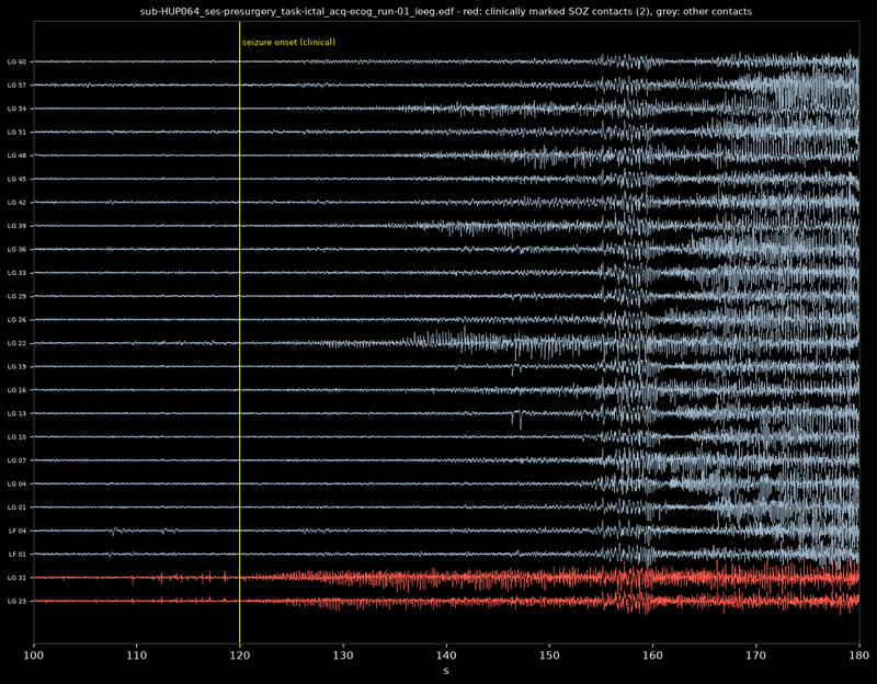
Beside the model: real patients. The HUP intracranial EEG dataset, 57 patients and 208 seizures with clinically marked onset channels and resections. In the app, HUP185's SEEG and HUP070's ECoG sit next to the simulation.
MethodsOpenNeuro ds004100 (HUP) · fitted Epileptor seizures hup-seizure-sub-hup185 and -hup070
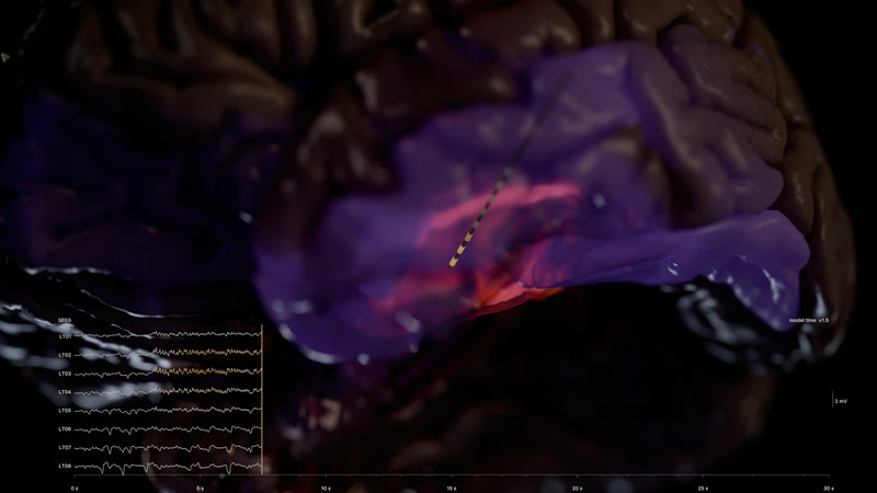
The seizure begins, under the depth electrode. 7.5 s into a simulated left mesial temporal seizure the parahippocampal cortex, the hippocampus and the amygdala light on BigBrain's own folds, seen from below and in front at 85 mm; the SEEG strip's contacts nearest the focus turn amber. The unlit hemisphere reads as dark tissue, the window is the brightest thing in the frame.
MethodsDelayed six-state Epileptor network on HCP-derived connectivity (dynamics/seizure-left-mesial-temporal-lateral-spread-distributed-v1: 82 regions, 35 s, OpenMEEG depth potentials), painted on bigbrain-cortex-hd; a modelled depth electrode and its 8 contacts' potentials in the strip · Blender 5.2 Cycles, frame 120, 1024 samples, 2560×1440 · simulated
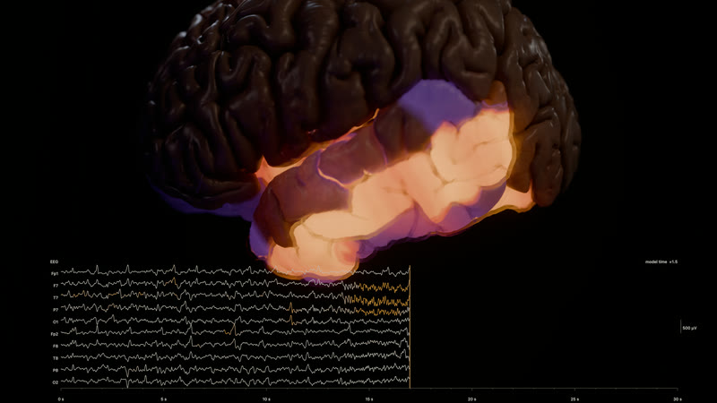
Recruitment: the temporal lobe lit at 17 s. The same seizure 17 s in, from the left: the superior and middle temporal gyri, the insula, the fusiform and the anterior cingulate are above a third of the model's activity range and the EEG's left temporal channels (F7, T7, P7) run amber. The run never crosses to the right hemisphere.
MethodsDelayed six-state Epileptor network on HCP-derived connectivity (dynamics/seizure-left-mesial-temporal-lateral-spread-distributed-v1: 82 regions, 35 s), painted on bigbrain-cortex-hd; scalp EEG from the run's MNE forward model in the strip · Blender 5.2 Cycles, frame 272, 1024 samples, 2560×1440 · simulated
Scene 5 · Neurons and microcircuits
Real human neurons, running.
One cell firing on its own reconstruction, a thousand-cell cortical circuit in health and depression, and the electron-microscopy tissue it all lives in.
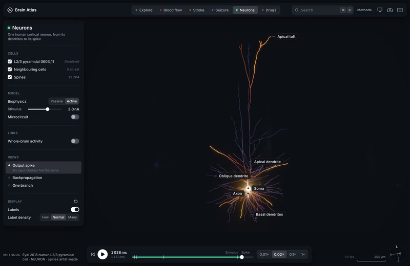
A human layer 2/3 pyramidal cell fires. Two back-propagating action potentials, a basal NMDA spike, then six synaptic clusters drive an output spike.
MethodsEyal 2018 cell 0603_11 in NEURON, its own reconstruction · run eyal2018-0603-11-bap-nmda-r2 · Blender 5.2 Cycles, 2560×1440
A living human microcircuit: 1,000 cells. Layer 2/3 at 34 °C. Firing rates PYR 1.0, SST 5.4, PV 10.1, VIP 3.4 Hz against the paper's 1.1, 5.6, 10.3 and 3.5. The EEG peaks at 11 Hz.
MethodsYao et al. 2022 human L2/3 microcircuit in NEURON + LFPy, 4.5 s with 2.5 s analysed, every compartment at 0.25 ms · run yao2022-l23net-baseline
The same circuit in depression. SST inhibition reduced by 40 %: pyramidal rates rise from 0.75 to 1.20 Hz and 5–16 Hz EEG power by 132 %.
A human layer 5 circuit. 100 cells at the paper's proportions at 34 °C; the 6.3 °C control stays within 5.9 % per cell type.
MethodsGuet-McCreight et al. 2022 human L5 microcircuit · run guetmccreight2022-l5net-test100
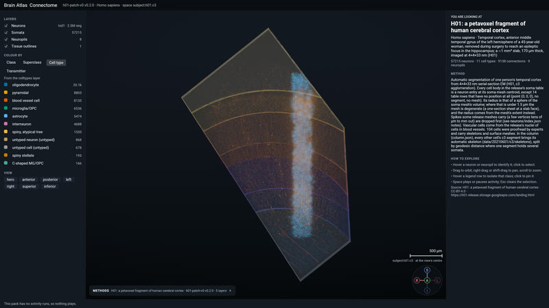
H01's column in the engine. 7,698 cell skeletons, 2,741 of them neurons, and 5.2 million synapse rows from a cubic millimetre of human temporal cortex, coloured by cell type.
MethodsH01 (Shapson-Coe et al. 2024), pack h01-patch-v0 0.2.0 · wgpu engine, 4K capture on M1 Max
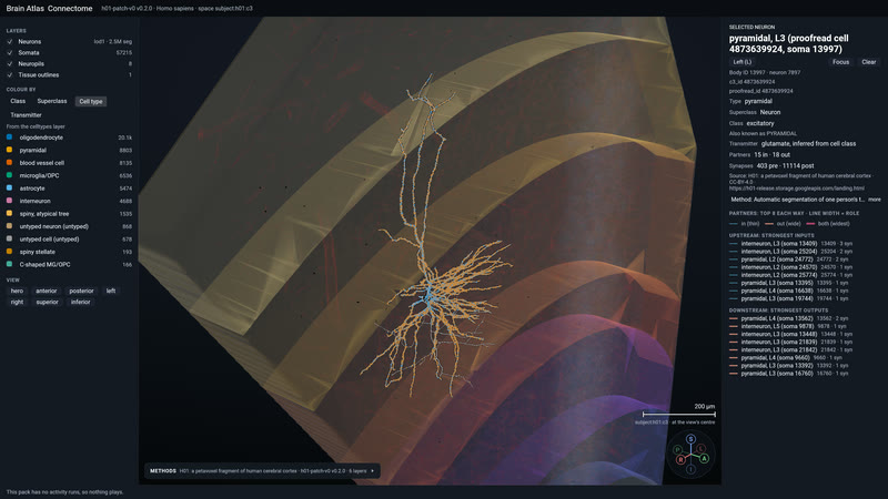
Cell 13997 and its synapses. One proofread cell selected with every synapse the electron microscopy found on it.
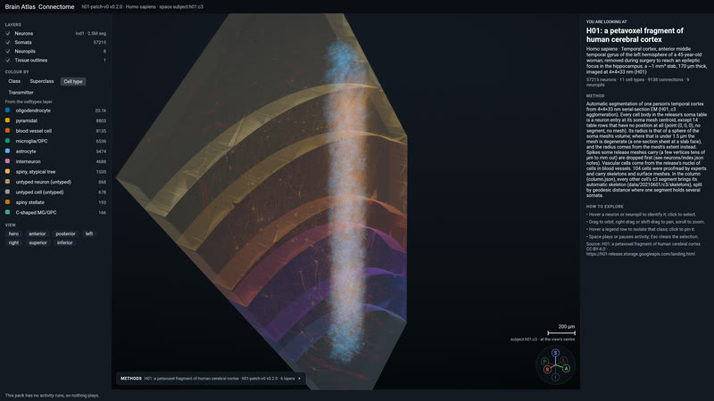
The whole cube, with its layers. 57,215 typed cell bodies from pia to white matter, with the cortical layers as films.
MethodsH01, pack h01-patch-v0 0.2.0, coloured by type · wgpu engine, anterior camera, 4K
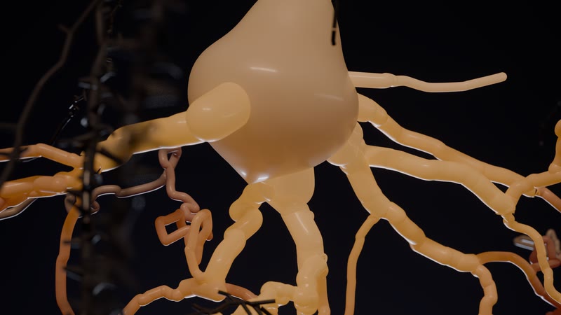
The soma under film light. A human layer 2/3 pyramidal cell glowing with its own simulated voltage, the 104 proofread H01 neighbours as ghosts behind it.
MethodsEyal 2018 cell 0603_11 in NEURON 9.0.2, synapse beads from run eyal2018-0603-11-bap-nmda · H01 proofread skeletons · Blender 5.2 Cycles, 768 samples, frame 96, 2560×1440
Look-dev: inside the cleft. Transmitter molecules crossing toward their receptors, at the molecular scale where the Powers of Ten zoom ends.
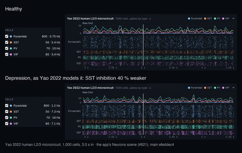
A human microcircuit fires, healthy and in depression. Yao 2022's human L2/3 circuit, 1,000 cells over 2.5 s, in the app's Neurons scene. Healthy, its pyramidal cells fire at 0.75 Hz, SST 5.4, PV 10, VIP 3.4. With SST inhibition 40 % weaker, the paper's depression condition, every type speeds up: pyramidal 1.2 Hz, SST 7.3, PV 16, VIP 7.1, 7,472 spikes against 4,618.
MethodsYao et al. 2022 human L2/3 cortical microcircuit (Cell Reports 38, 110232) in NEURON 8.2.7 at 34 °C: 800 pyramidal, 50 SST, 70 PV and 80 VIP cells on replicated Allen human morphologies; runs cells/yao2022-l23net-baseline and cells/yao2022-l23net-depression, depression the paper's MDD condition with SST synaptic and tonic inhibition reduced by 40 %; rates and rasters are each run's own somatic spikes over the recorded 2.0–4.5 s (metrics.json: pyramidal 0.7525 against 1.2005 Hz; the paper reports 0.77 and 1.2); shown by the app's Neurons Model → Microcircuit at 3.0 s, the circuit not yet drawn in the 3D view · simulated
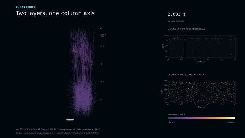
Two layers, one column axis. Yao 2022's human layer 2/3 circuit (20 recorded cells) sits over Guet-McCreight 2022's layer 5 (100 cells) on a shared pia axis, as voltage-coloured morphologies with their rasters. Two independent NEURON baselines aligned in the H01 frame, not yet a coupled column: the coupled run is the next bet.
MethodsRuns yao2022-l23net-baseline-closeup-r2 and guetmccreight2022-l5net-test100-r2 (NEURON), frames at 2.632 s, placed by rigid translation in the h01-patch-v0 0.2.1 column frame · derived
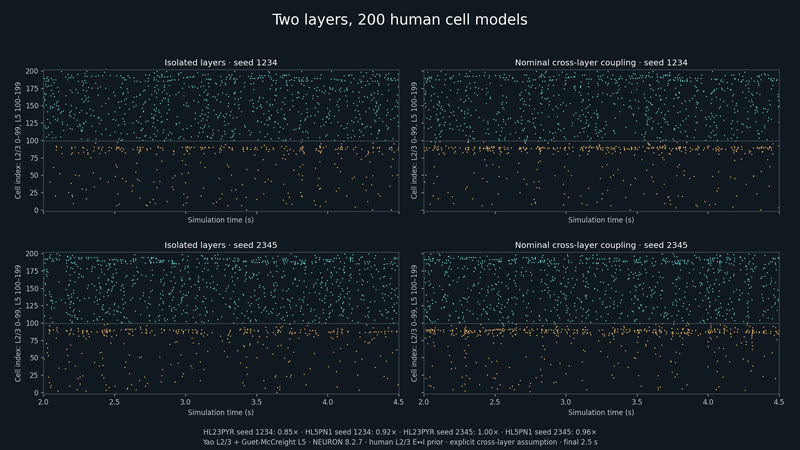
Two layers, 200 human cell models. Nominal coupling raises L2/3 PV spike counts 1.73× and 1.71× in two simulated seeds.
MethodsYao L2/3 and Guet-McCreight L5, 100 cells each in NEURON 8.2.7; source OU input, human-informed within-L2/3 E↔I probabilities, assumed Potjans–Diesmann cross-layer wiring, retained uniform synapse RNG. Raster: final 2.5 s of 4.5 s runs, human-isolated versus human-coupled, seeds 1234 and 2345 (ACT-002). · simulated
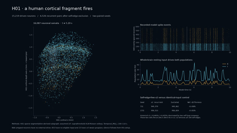
Human cells, whole-brain input. 15,219 driven neurons fire in 10-second simulations; removing 104 self-edges eliminates the runaway cells.
MethodsH01 soma locations and sparse segmentation-derived subgraph; assumed exponential-current LIF with suprathreshold Poisson volleys from ALN Temporal_Mid_L, 100–110 s. Post-hoc self-edge-free v2, 6,526 recurrent pairs, two identical-input controls. Untyped neurons have no external drive; 853 have no eligible input and 15 have at most five weak synapses. Pack h01-patch-v0 0.3.0, default h01-lif-resting-recurrent-seed731-v2. · simulated
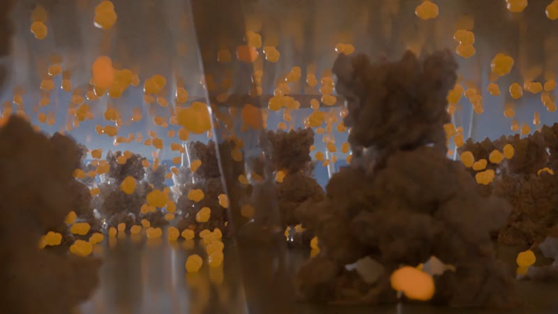
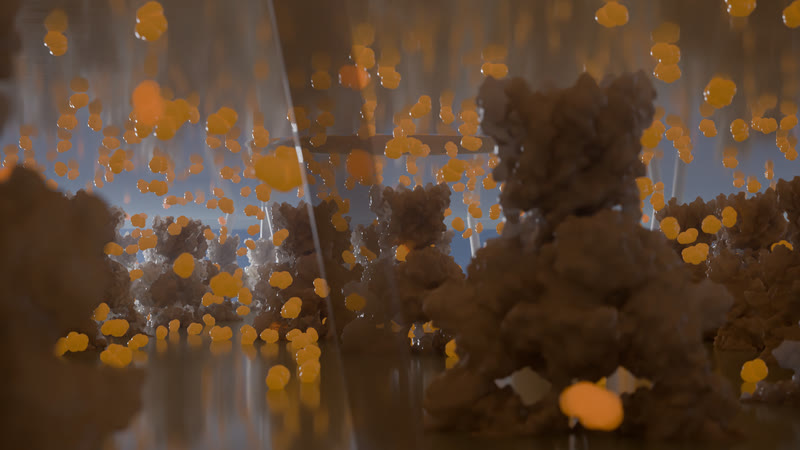
Inside the synaptic cleft. Glutamate crossing the 20 nm cleft 100 microseconds after release: a burst of 1,500 molecules from one fused vesicle, binding AMPA and NMDA receptors on the spine. Frame 430 of the neurons sequence, 2560×1440.
MethodsMolecule positions from the Smoldyn run synapse-glutamate-nbqx-zero-drug (1,500 glutamate every 20 µs for 43 ms in a 300 nm × 20 nm cleft, 80 AMPA + 20 NMDA sites with kinetic states), drawn where the run puts them; receptors are PDB 3KG2 (GluA2) and 7SAA (GluN1/GluN2B) as Molecular Nodes surfaces at the run's sites; the bouton, vesicle, pore, spine and membranes are modelled from literature dimensions. Cycles, 768 samples, 85 mm at f/0.35, the neurons scene at 1 nm = 1 m. · simulated
Scene 6 · Drugs
Dose, then blood, then brain.
Receptors bind by real densities and affinities across the cortex, and at the synapse, molecules meet their receptors one at a time.
1,500 glutamate molecules, with and without the blocker. A 43 ms synaptic event as particles: NBQX competes for the AMPA receptors and cuts their integrated open time by 87 %.
MethodsSmoldyn particle simulation, Jonas AMPA and Erreger NMDA kinetics, 80 AMPA and 20 NMDA receptors on a 300 × 300 nm face · 12-seed ensemble: 87.45 % lower AMPA open time · run synapse-glutamate-nbqx
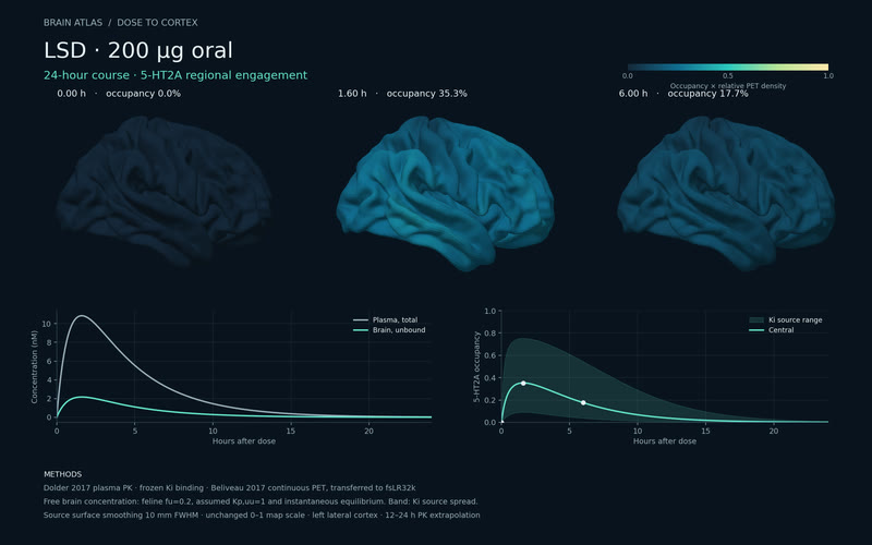
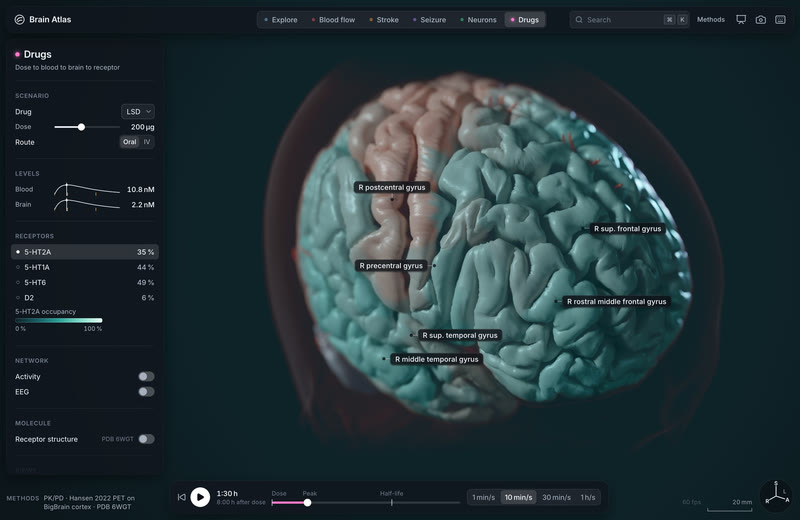
LSD, 200 µg, over 24 hours. Plasma, free brain concentration and 5-HT2A occupancy, peaking at 35 % at 1.6 h, painted on the cortex by receptor density.
MethodsDolder 2017 oral PK, frozen Ki binding, Beliveau 2017 5-HT2A PET map on fsLR · run lsd-200ug-cortex-v1
5-HT2A engagement, 96 minutes after 200 µg of LSD. Occupancy times PET receptor density, painted on the template cortex under film light.
MethodsDolder 2017 oral PK and equilibrium site binding, Hansen 2022 5-HT2A density · MNI152 2009c pial surface · Blender 5.2 Cycles, 512 samples, 2560×1440
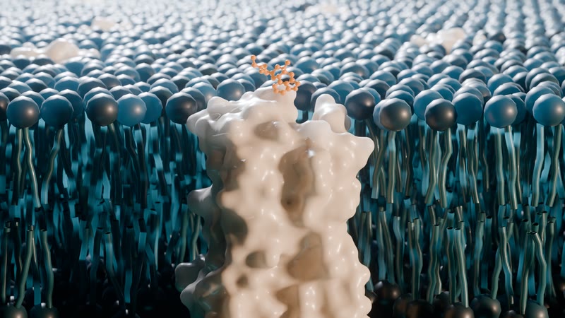
LSD seated in its receptor. The 5-HT2A receptor cut open in its lipid bilayer, the drug's 24 heavy atoms in the crystal pose as the receptor shifts to its drug-bound state; at the peak, 45 of 127 receptors carry it.
MethodsPDB 6WGT (LSD-bound 5-HT2A) morphing to 9WPQ, Molecular Nodes; occupancy from the PK course, 96 min after 200 µg · Blender 5.2 Cycles, frame 241, 512 samples, 2560×1440
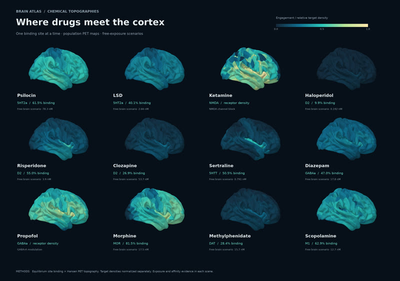
Where 12 drugs meet the cortex. One binding site at a time: 19 PET targets, 2,579 measured affinities, free-exposure scenarios.
MethodsEquilibrium site binding × Hansen 2022 PET maps · pharma atlas, 19 targets on fsLR
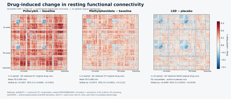
Measured, not modelled: how three drugs change connectivity. Global shift in Fisher z: psilocybin +0.107, methylphenidate +0.057, LSD +0.010. Global-signal regression flips the signs. The receptor-informed model is the next step.
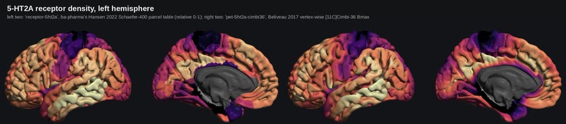
5-HT2A density on the HD cortex. The receptor map the drug scenes paint with: the Schaefer-400 parcel table on the left, the vertex-wise PET map on the right.
MethodsHansen 2022 parcel tables and Beliveau 2017 vertex-wise 5-HT2A PET, on the cortex-hd pack
The app
Six scenes, one engine, in your browser.
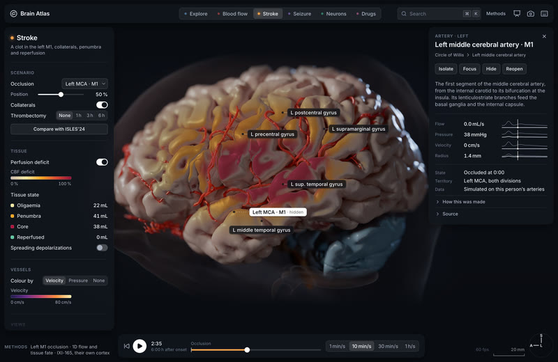
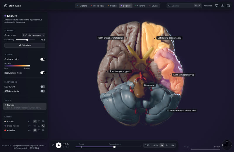
A web app on the wgpu engine, built for WebGPU: Explore, Blood flow, Stroke, Seizure, Neurons and Drugs, each with a Methods panel naming its model and data. Free to use.
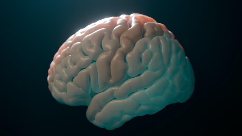
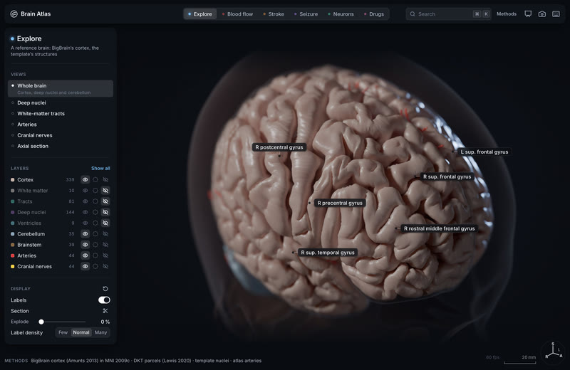
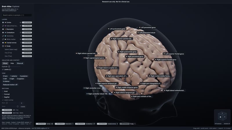
Explore in the native engine. The template's cortex, deep nuclei and cerebellum in the default load state: reference anatomy, an average of 152 adults.
Methodsmni152-2009c-alpha pack: FastSurfer segmentation of the MNI152 2009c template · wgpu engine, native capture, hero camera
The same brain in Chrome. The engine's WebGPU/wasm build, which the app hosts.
Methodswgpu engine, wasm build · Chrome WebGPU at 1600×960 @2×
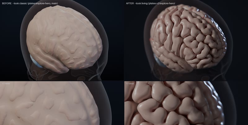
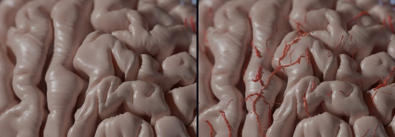
The living look: before and after. Design's plates moved from the classic model look to living tissue: subsurface, a wet sheen, veins in the sulci.
Methodsart-direction notes, the mockup plates in the living look (2026-10-01) · Blender 5.2 Cycles
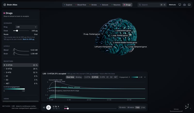
The dose is a slider: LSD's course re-fit live at 100 µg. Slide the dose from the course's 200 µg to 100 µg and the whole course re-fits on its own model: 5.42 nM in blood and 1.08 nM in brain at 1 h 36, 5-HT2A 21 % occupied, 5-HT1A 28 %, 5-HT6 32 %, D2 3 %.
MethodsDrugs: pharma/lsd-200ug-cortex-v1 (oral one-compartment apparent PK, dose to continuous cortex), rescaled live to 100 µg oral on its own model and read at 1 h 36; receptor occupancy from each receptor's Ki range (band on the 5-HT2A trace); drawn by the wgpu engine 42fa1136 (Chrome, 2x) · simulated
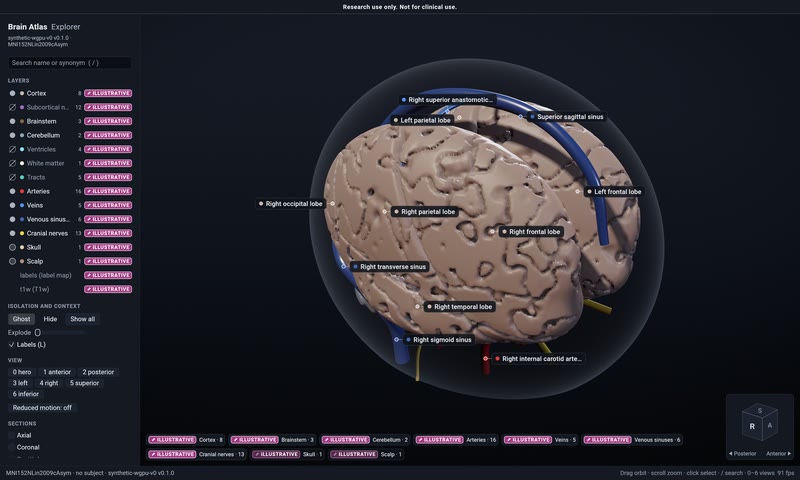
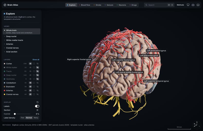
Explore opens on BigBrain's cortex. The app's first scene is now a reference brain: BigBrain's cortex, one person's brain rebuilt from 7,404 histological sections at 20 µm and placed in MNI 2009c, with the template's arteries, cranial nerves, cerebellum and brainstem around it, drawn live in Chrome's WebGPU viewport.
MethodsBigBrain cortex (Amunts et al. 2013), placed in MNI152 2009c by an affine, with DKT parcels per vertex (Lewis et al. 2020); around it mni152-2009c-v0's template structures: FastSurfer's nuclei, cerebellum and brainstem, the Mouches and Forkert arterial atlas (2019) and Z-Anatomy's cranial nerves. Drawn by the wgpu engine 9f964b6 (Chrome, 2x) · reference
H01's real wiring, firing: a cubic millimetre of human cortex in the app. Neurons' Its tissue plays H01's own default run on its 10 s clock: 16,087 neurons of one person's temporal cortex, wired by the 6,526 connections segmented from the electron microscopy, spiking warm through the column's six layers under a resting background drive. The 15,219 driven neurons aren't ringed or flashed cool, so what lights is the tissue itself.
MethodsH01 (Shapson-Coe, Januszewski, Berger et al., Science 2024; Lichtman Lab and Google Research): h01-patch-v0 0.3.0, run h01-lif-resting-recurrent-seed731-v2: exponential-current LIF with phenomenological parameters on H01's eligible neuronal graph, 6,526 of 9,138 connections kept (9,113 synapses; the 104 self-edges excluded), independent Poisson afferent volleys at ALN Temporal_Mid_L E/I rates into 15,219 of 16,087 modelled neurons, seed 731, 949,579 spikes over 10 s; at 4.0 s, drawn by the wgpu engine 42fa1136 (Chrome, 2x) · simulated
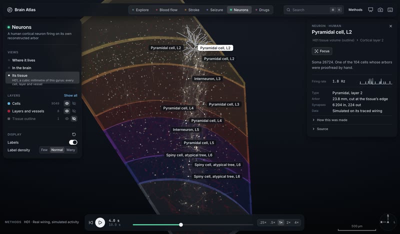
One neuron of H01, read from its own tables: the app's cell inspector. Select any cell in Neurons' Its tissue and the inspector says what it is and how it fired in the run: the column's centre pyramidal cell in layer 2 fires at 1.8 Hz (18 spikes in the run's 10 s), over its rate in each 100 ms, with 23.8 mm of traced arbor that runs out of the imaged tissue and 6,204 synapses in and 224 out. Glia have no rate, and a neuron the run never drives reads Silent.
MethodsH01 (Shapson-Coe, Januszewski, Berger et al., Science 2024; Lichtman Lab and Google Research): h01-patch-v0 0.3.0's neuron table (cell types, 2024 layers, skeleton cable, cut flags, synapse rows) and its default run h01-lif-resting-recurrent-seed731-v2 (exponential-current LIF on H01's eligible neuronal graph, Poisson afferent volleys, seed 731), read by the app from the run's rates.u8, which match its spike counts exactly; at 4.0 s, drawn by the wgpu engine 42fa1136 (Chrome, 2x) · simulated
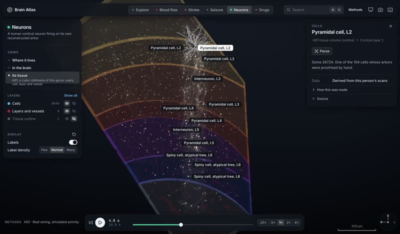
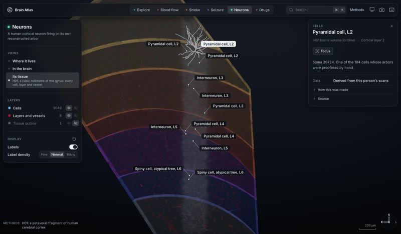
Down to the tissue: H01's cubic millimetre in the app. Neurons' new view opens H01, a cubic millimetre of one person's temporal cortex reconstructed by electron microscopy, in the app's WebGPU viewport: 9,049 named cells (the 104 whose arbors were proofread by hand and the column's 8,945 others), six cortical layers, the white matter and the vessels, with layer-2 pyramidal cell 26724 picked out.
MethodsH01 (Shapson-Coe, Januszewski, Berger et al., Science 2024; Lichtman Lab and Google Research): 4×4×33 nm serial-section EM, c3 agglomeration, 104 cells proofread. Drawn from h01-patch-v0 0.2.1 (neuron table, proofread skeletons, layer and vessel meshes) by the wgpu engine 9f964b6 (Chrome, 2x) · derived
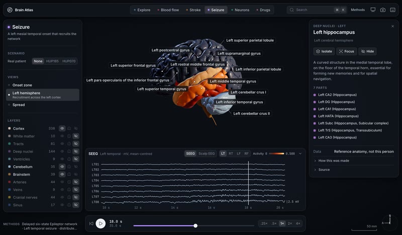
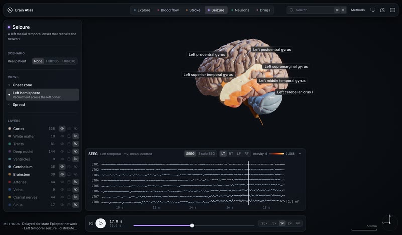
A seizure lights BigBrain's own cortex, live in the app. 17 s into a simulated left mesial temporal seizure, 9 of the model's 82 regions are above a third of its activity range, and the app's WebGPU viewport lights them on BigBrain's real folds (163,842 vertices per hemisphere): the superior temporal gyrus at 98 %, the insula at 74 % and the middle temporal gyrus at 69 %, with the depth electrodes' discharges in the strip below.
MethodsDelayed six-state Epileptor network on HCP-derived connectivity (dynamics/seizure-left-mesial-temporal-lateral-spread-distributed-v1: fsaverage, 82 regions, 1,051 frames over 35 s, OpenMEEG depth potentials), moved onto bigbrain-cortex-hd 0.1.0 through its map-fsaverage by the wgpu engine 9f964b6 (Chrome, 2x) · simulated
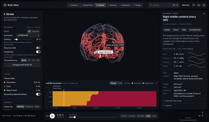
Any vessel, live: its flow, its territory, and Occlude here. Two and a half hours into a left M1 clot, pick the other M1 and the inspector reads it from the live solve: 1.85 mL/s at 91.9 mmHg and 28.7 cm/s through a 1.43 mm radius, open, feeding the right MCA territory, with Occlude here to put the clot there instead and re-solve the whole network.
MethodsStroke at 2 h 35 after a left M1 occlusion: tissue hemo/ixi165-stroke-v5-l-m1-intermediate-untreated, flow hemo/ixi165-1d-v5-occl-l-m1 with hemo/ixi165-1d-v5-baseline as the reference, spreading depolarizations dynamics/spreading-depolarization-ixi165-m1-v5-841dcfd0-t0-v1; the live rows are hemo-rt's cycle means on the v5 Circle of Willis network (a model circle, not this person's), the territory is each region's share of its blood through this vessel by the reference run's mean flows, mixing fully at each junction; Occlude here moves the clot and re-solves; drawn by the wgpu engine 42fa1136 (Chrome, 2x) · simulated
Every bet writes its kill rule before the data are opened, and the kills stay on the page. So far: geometry and metabolism hold at the map level, the physics checks out, and template networks at n ≈ 50 can't carry mechanistic claims.
Perforator territory, distance from the watershed and large-artery probability predict lesion frequency. The high-extraction mechanism does not (p = 0.916).
MethodsISLES'22 lesion-frequency map (400 cases) × Liu 2023 territories, the Mouches–Forkert artery atlas and neuromaps CBF and CMRO2; BrainSMASH spatial nulls · bet 2
Kept
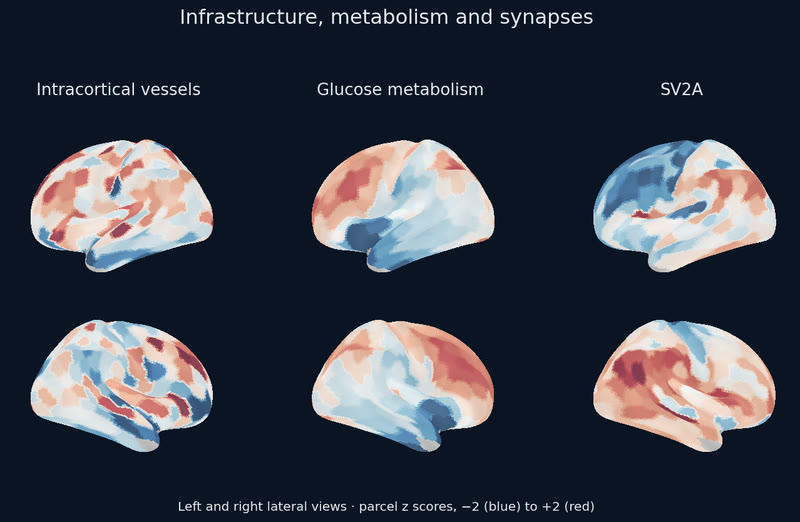
Cortical blood flow tracks glucose use, not synapse density.
Partial r 0.51 (ASL) and 0.80 (PET) with CMRGlu; 0.30 and 0.02 with SV2A.
394 Schaefer parcels, controlling cell staining. The strong synaptic form of the hypothesis is opposed by the intracortical vessel map.
Methodsneuromaps CBF, CMRGlu and CMRO2; UCB-J SV2A; BigBrain staining; spin tests · bet 5
Killed
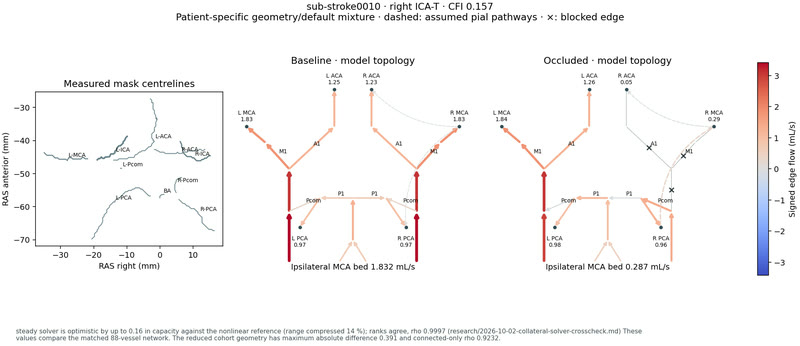
A patient's Circle of Willis physics predicts infarct growth.
ΔR² −0.016 [−0.071, 0.005] for growth over the clinical baseline.
48 strict anterior-circulation ISLES'24 cases in nested cross-validation: the capacity index does no better than a 4-bit variant code. The physics, the 149-case feature table and the site inventory stand.
Methods1D steady solver on each patient's TopCoW circle graph, cross-checked against openBF (rank ρ 0.9997) · bet 1
Killed
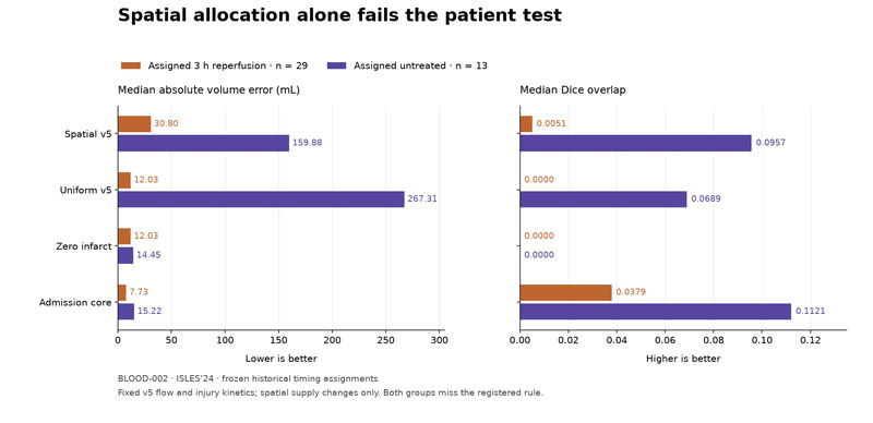
Spatial supply alone predicts a patient's final infarct.
Median volume error 30.8 mL, against 12.0 for zero infarct and 7.7 for the admission core (3 h group, n = 29).
Untreated, n = 13: 159.9 mL against 14.5 and 15.2. IXI-165's v5 supply routed into 42 ISLES'24 cases at fixed injury kinetics, the rule frozen before scoring. v5 itself met its registered rule, and predicting no infarct at all still beat it: 12.7 mL against 37.0.
MethodsPassive route tracing of the v5 flows into each case's own vessel map, 29 assigned 3 h and 13 untreated, scored natively against the ISLES'24 final infarct with uniform, zero and admission-core controls · bet BLOOD-002, research note 2026-10-02-blood-spatial-result
Killed
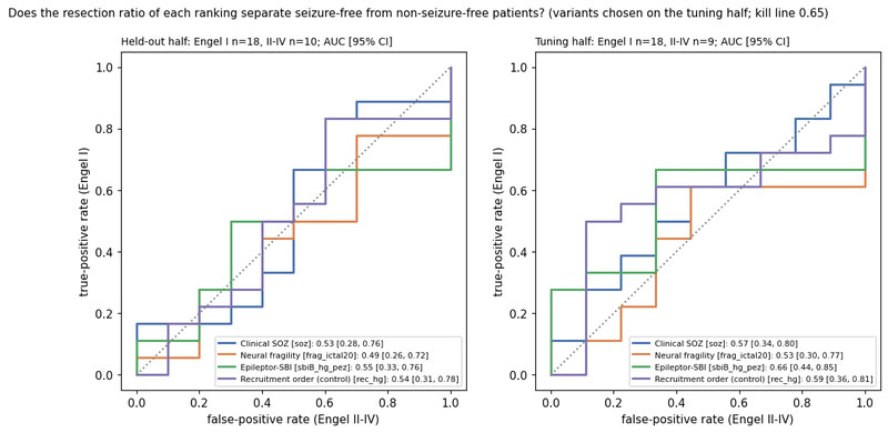
A template Epileptor fitted by simulation-based inference finds the seizure focus.
Held-out AUC 0.55 [0.33, 0.76] against a 0.65 bar.
Neural fragility 0.49, the clinical onset zone 0.53, recruitment order 0.54. Every ranking finds the resected channels (AUC 0.68–0.74); none predicts the outcome in this cohort.
MethodsHUP ds004100, 55 patients split into tune and held-out halves, Engel I vs II–IV · bet 7
Killed
Seizures spread along white matter, not across the cortex.
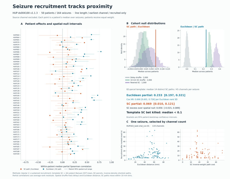
50 patients, 164 seizures: recruitment order follows proximity, with a hazard ratio of 0.666 per distance-rank SD. Patient-specific tractography is untested.
MethodsHUP ds004100 recruitment times × an 82-parcel template connectome; registered plan · bet 9
Won
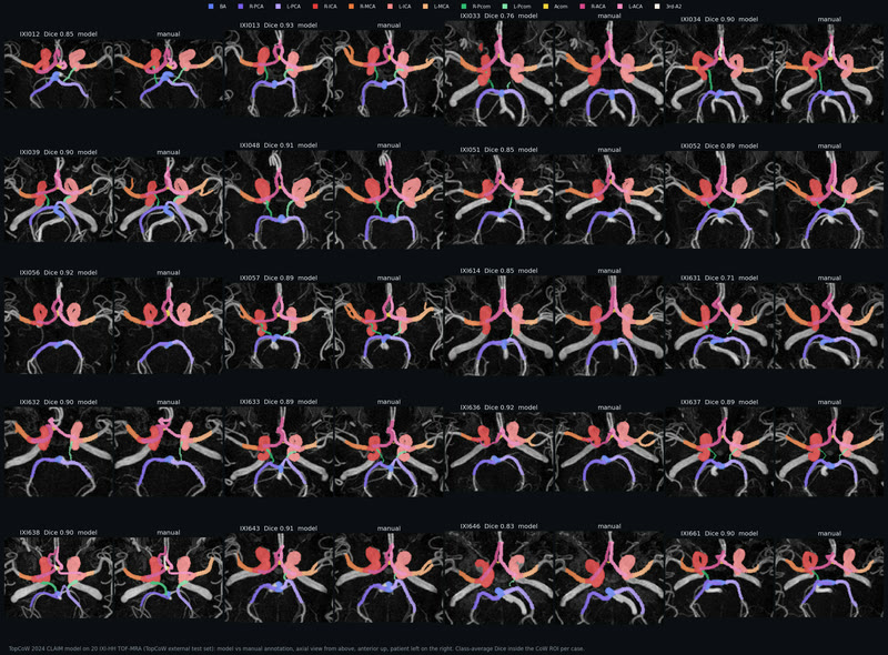
Our Circle of Willis segmentation runs on our own GPU.
Class Dice 0.876, merged-vessel Dice 0.960, clDice 0.982 on 20 fresh IXI brains.
27.7 s a brain against 2 min 50 s in the reference Docker, reproducing it to one voxel. The small communicating arteries carry almost all the error.
MethodsTopCoW 2024 nnU-Net weights, IXI-HH TOF-MRA against manual annotation
Won
The clot's position along M1 decides the deep infarct.
Lenticulostriate bed lost: 30 % proximal vs 13 % distal (p = 0.0026).
In 72 ISLES'24 M1 patients the loss falls with distance from the carotid terminus (ρ = −0.37). Insula and cortex don't differ, so the effect is specific to the end-artery bed.
MethodsISLES'24 training set, clot near-edge measured on CTA · bet ISLES-003
Killed
A learned surrogate makes 1D cerebral flow real-time.
22.7 % held-out flow error, 13× faster than physics that already solves in 0.85 ms.
The MLP buys no interactivity on this graph: the exact periodic solver already answers in the browser in 1.7 ms. Nonlinear teachers are next.
Methodsa learned MLP surrogate against the exact Rust/Wasm periodic solver hemo-rt
Won
The engine's cortex matches the reel at its own camera.
Luma quartiles within 0.02 of the Cycles still.
IQR 0.12 to 0.37 against the reel's 0.36; the top 1 % is still short. Killed alongside: the capillary net as colour, which reads as a red honeycomb on the engine.
Methodsengine look bets ENGINE-LOOK-001 and -002
Measured
Three drugs, three connectivity fingerprints.
Psilocybin +0.107, methylphenidate +0.057, LSD +0.010 global Δz.
35 unique acquisitions from seven people scanned repeatedly, plus 15 LSD participants. The targets for the receptor-informed model are published; the model stage is next.
Methodsds006072 and ds003059, pre-registered plan frozen before the FC computation · bet 3
Data
Real data at every scale.
55 datasets requested, 46 acquired and verified, each with its own manifest saying where it is and how to fetch it again.
264dataset manifests, each saying where the data is and how to fetch it again
46of 55datasets acquired and verified; the rest wait on access approvals
2.7TBof raw data on the project's store
7,404stained sections in BigBrain, one human brain at 20 µm
503,085streamlines in the HCP1065 tract atlas, 102 M points before simplification
163,842vertices a hemisphere on the canonical cortex, BigBrain's own
57,215typed cell bodies in the H01 pack; its column holds 7,698 skeletons and 21.0 M synapse points
149+ 250stroke cases on disk from ISLES'24 and ISLES'22, plus 1,452 strokes in ATLAS v3.0
57HUP intracranial EEG patients, 208 seizures
19PET receptor targets and 2,579 measured affinities behind the drug scenes
35+ 60rest-fMRI runs behind the drug fingerprints: psilocybin and methylphenidate (ds006072), LSD (ds003059)
1mm³of human cortex in H01 at 4 × 4 × 33 nm, 6.1 GB of release subset on disk
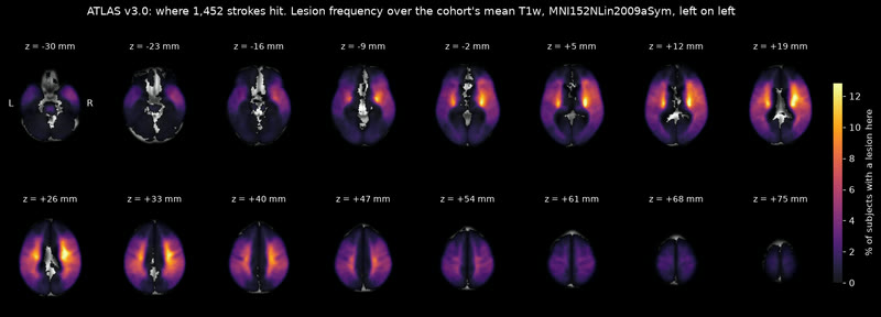
Where 1,452 strokes hit. ATLAS v3.0 lesion frequency; the peak is 15.5 % of subjects, in the right putamen.
MethodsATLAS v3.0 lesion masks, voxel-wise frequency over its 1,452 subjects
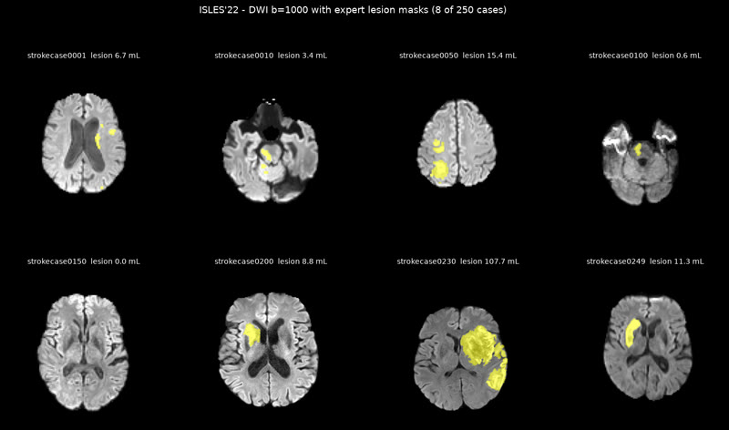
ISLES'22: 250 cases with expert lesion masks. Eight of them on diffusion MRI; the challenge's 400-case aggregate heatmap is what the lesion-geography bet used.
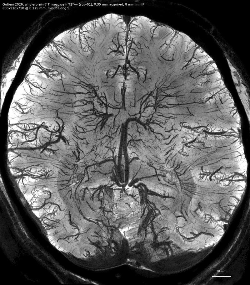
Whole-brain veins at 7 T. The mesoscale venous map that will drain the living brain.
MethodsGulban 2026 meso-vein 7 T
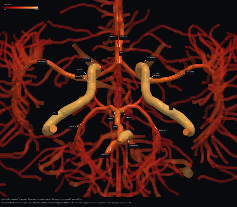
The named circle. vessels-hd: 729 arterial segments (10.4 m of vessel) and 612 venous (7.1 m) on the template, the Circle of Willis labelled, seen from below.
Methodsmni152-2009c-vessels-hd 0.1.1, decoded from the pack's graphs
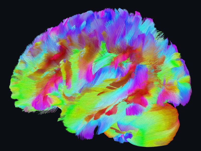
The dense tract pack, lateral view. All of HCP1065 as tiers the browser can stream.
Methodsmni152-2009c-tracts-dense: RDP at 0.1 mm, u16 quantised chunks in tiers of 1/16, 1/4 and all
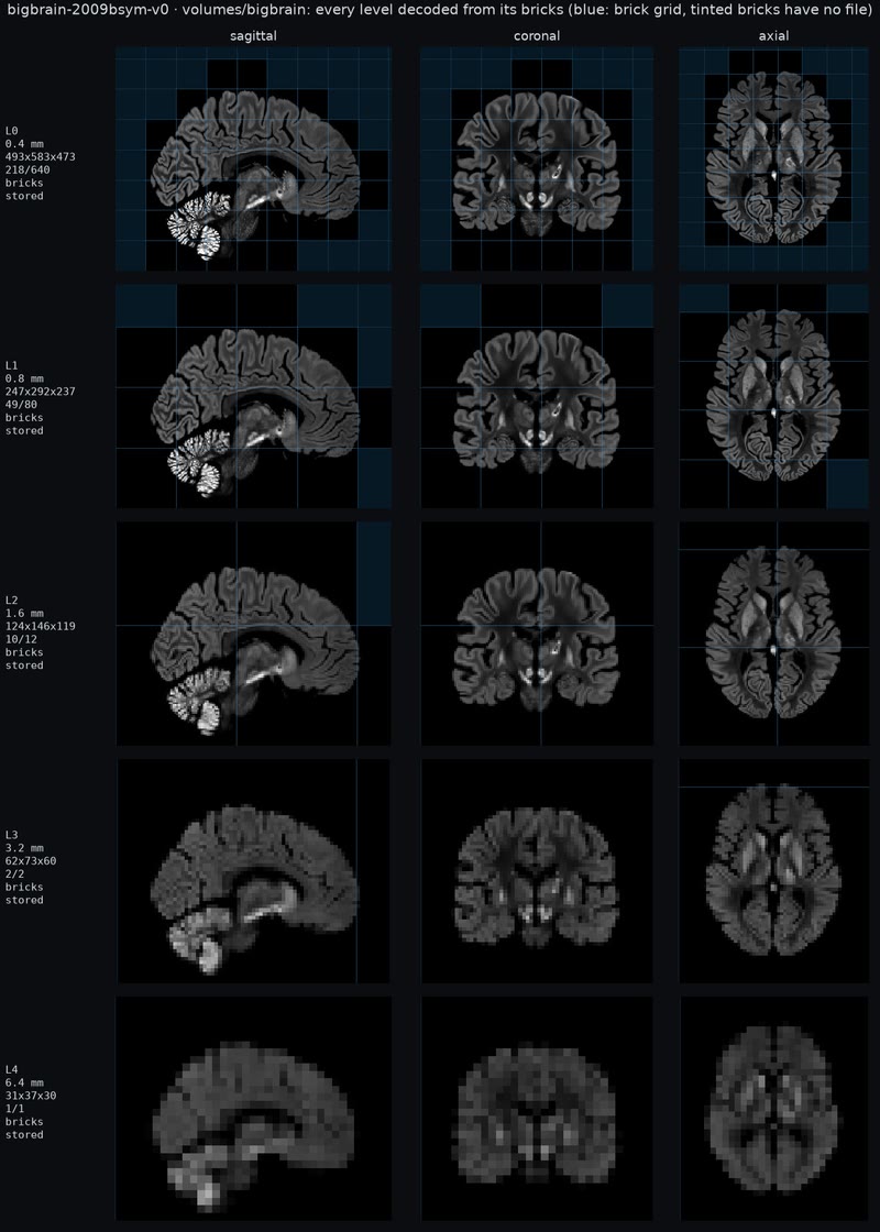
BigBrain's brick pyramid. Every level decoded from its own bricks, from 6.4 mm down to 0.4 mm, in three planes: what the volume renderer streams.
Methodsbigbrain-2009bsym-v0 volume pack from BigBrain 2015; 218 of 640 bricks stored at the finest level
Real pial arteries on BigBrain's cortex. The same crop of the Explore hero without and with the 7 T pial arteries (Bollmann 2022) draped onto BigBrain's folds: 26 m of real vessels in one network, now a published pack.
MethodsThree 7 T time-of-flight acquisitions (Subject_03, Subject_01, twoEchoTOF) fitted to BigBrain's pial surface in MNI 2009c by a trimmed iterative closest point, every centreline point moved to the nearest surface point and lifted by its radius, the three feathered into one network by leaf pruning. A drape, not a registration: the vessels are real, their course on these folds is not. Cycles, living look, 2940×1912, 256 spp. · derived