From the whole head to a single synapse: real anatomy, real physics and real data at every scale, rendered at cinematic quality. Free and open, for research and education.
The app needs a WebGPU browser: desktop Chrome or Edge, or recent Safari.
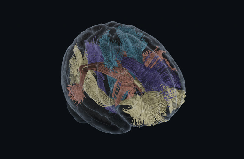
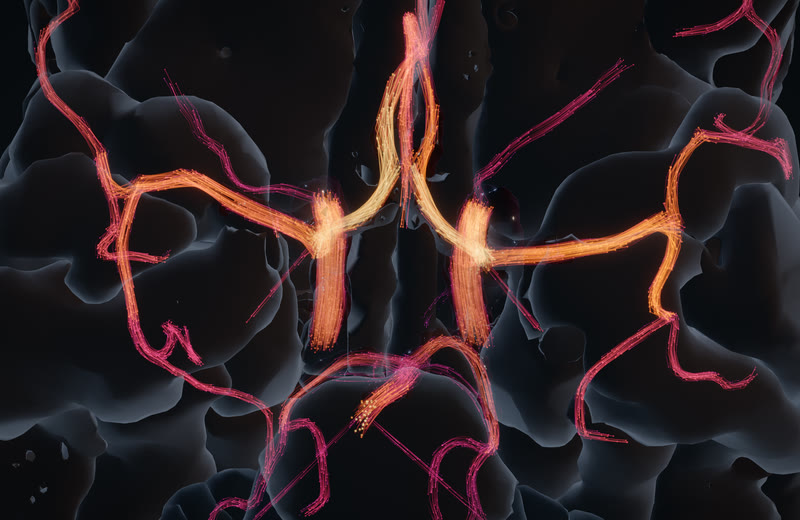
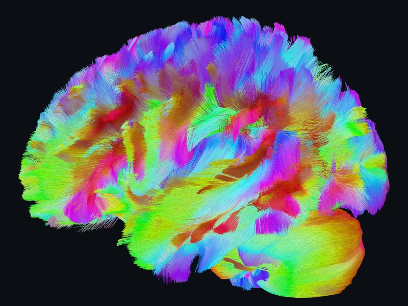
MethodsHCP1065 population tractography (Yeh 2022), 503,085 streamlines resident and drawn as lit tubes with fibre-scale occlusion under a distance LOD, on the MNI152 2009c cortex · wgpu engine, native 2940×1912 capture, M1 Max · reference
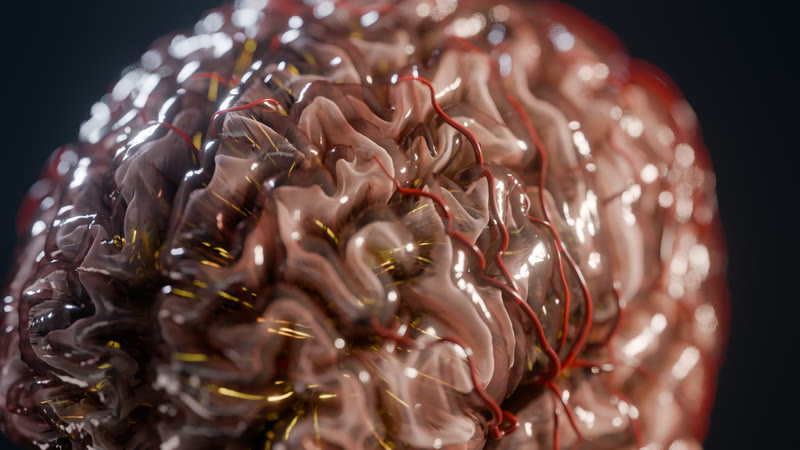
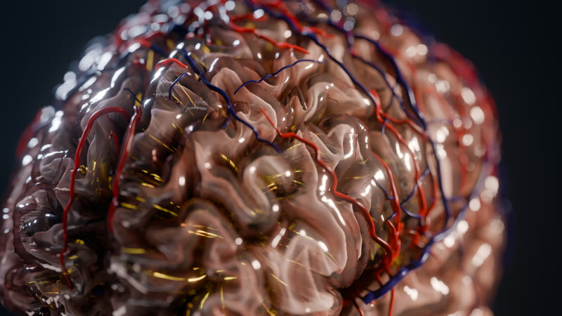
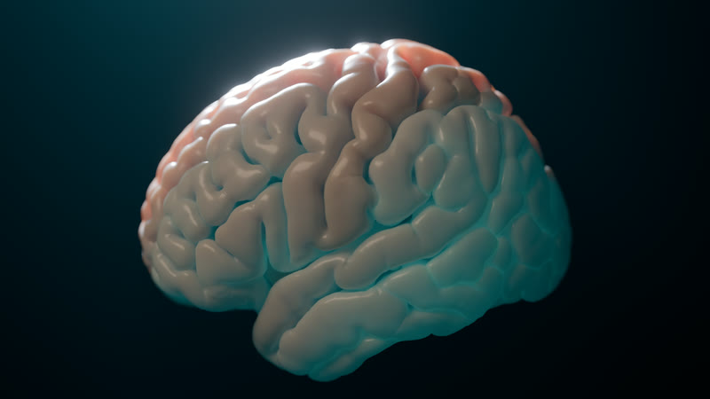
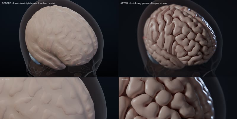
MethodsThe Living Brain, hero v2 under design's living look (sulc-percentile colour, 6 W/m² key 70° off axis, 4350 K) · BigBrain's own cortex (Amunts et al. 2013) turning translucent over the HCP1065 fibre bundles, arteries pulsing with the 1D model · Blender 5.2 Cycles, 2560×1440 · reference
The Wave 1 reel, 1 October 2026.
MethodsThe Wave 1 reel, 1 October 2026: a cut of the gallery’s films and stills; each shot’s model and data are in the Methods line of its own card · simulated
The 90-second reel, rough cut v7. From the whole head to one receptor, with music: the Living Brain hero opens it, then blood flow through IXI 165’s own arteries in the app’s engine, the clot and the 12 h territory, the seizure’s onset deep in the left temporal lobe spreading under its SEEG strip, the tissue into the firing cell, the synapse and glutamate crossing its cleft live on the GPU, LSD seating in its receptor, the TMS coil orbiting C3 in the app, and one continuous dive from the head to a single receptor. Tap to watch.
MethodsCut from hero-v2 (look v2); the blood-flow, seizure, neurons and drug-binding finals (Cycles, 1920×1080, 128 spp) trimmed to their beats; three pieces from the app’s engine: 3D flow through IXI 165’s arteries with wall shear stress and 20,000 tracers (7 s), the TMS coil’s 5 s camera orbit over C3 on IXI 165 from the live app, and the Powers of Ten dive from dive-head 0.3.0 at 48 steps a second, at the journey’s pace then twice it to the receptor at 30 s, with a scale readout; the living synapse, a glutamate Monte Carlo run live on the GPU (the e2-cleft-live card’s clip); caption cards from the render sidecars; music by Eleven Music at −16 LUFS; 0.5 s dissolves · 17 segments, 1920×1080, 24 fps, 95.5 s · simulated
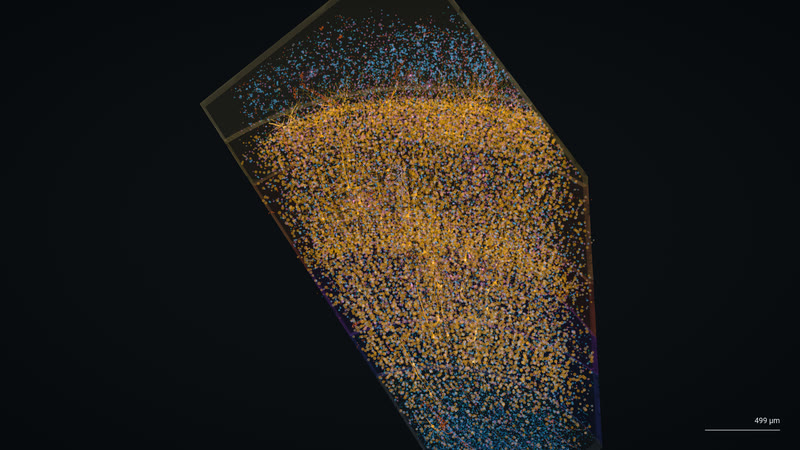
A whole cortex, then BigBrain's stained sections at 100 µm, then H01's electron-microscopy neurons: one push, no cuts, every pixel from a measured dataset.
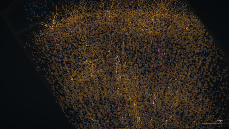
From the cortex to one neuron in an 18-second push. The pial surface fades as the camera magnifies into the 100 µm cut, BigBrain's own Merker stain, which dissolves into H01's column of 7,698 reconstructed cells, 2,741 of them neurons, and settles on a single proofread cell.
Methodswgpu engine 6d99a0af (Powers of Ten v2), native Metal capture at 1080p30, the pial fading as it magnifies and the cut edge inked · BigBrain 2015 (Amunts et al. 2013) at 100 µm, ray-marched bricks in the Merker stain · H01 human temporal cortex EM, pack h01-patch-v0 0.2.1: 7,698 skeletons, 5.2 M synapse rows · measured
Every bundle in the atlas. 503,085 streamlines resident in four anatomical colour families, drawn as lit tubes with fibre-scale occlusion, depth fog and a distance LOD.
MethodsHCP1065 (Yeh 2022), 91 bundles simplified to 15.5 M points at 0.1 mm · wgpu engine, 2940×1912 · reference
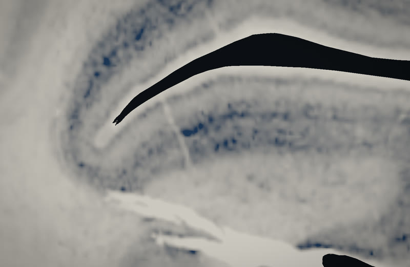
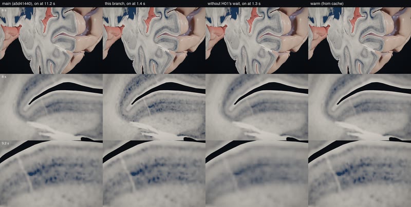
Powers of Ten, first visit. On a cold first visit the dive now starts in about 2 s, where it waited 11 s, and the 100 µm histology is in as the camera arrives.
MethodsBigBrain 100 µm histology (Amunts et al. 2013) on the Powers of Ten path, 8.0 s into the 18 s dive. Headless Chrome, 1470×956, cold cache, the public app with engine cedd5fa2. Interleaved with engine a5d41440 on one link: journey on in 1.3–1.9 s vs 10.1–11.2 s; stills at 6, 8 and 9.2 s as close to a cached play as before, or closer. · measured
Cold start against the cached play. The same three moments of the dive on a cold first visit, before and after, beside a play from the browser's cache: the stills now match, and the dive starts about 9 s sooner.
MethodsA/B stills of the Powers of Ten dive at 6, 8 and 9.2 s on a cold first visit to the public app: the previous embed (a5d41440, on at 11.2 s), the new one (cedd5fa2, on at 1.4 s in this run; about 2 s since: 1.4–3.5 s over batches 59–61's live checks and an 8-pair interleaved A/B of batches 60 and 61, medians 2.25 and 2.21 s), without H01's wait (1.3 s), and a play from the browser's cache. Headless Chrome, 1470×956, interleaved runs, 2026-10-03. Mean per-pixel |Δ| from the cached play at 8 and 9.2 s: 0.0/0.0 before, 0.2/0.0 after. · measured
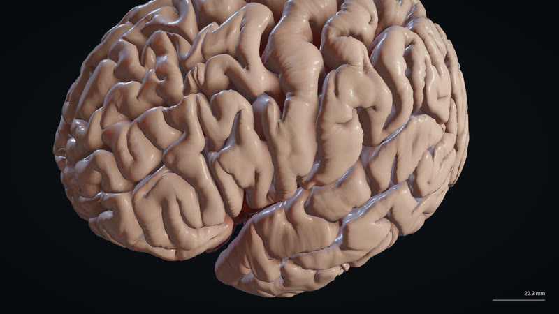
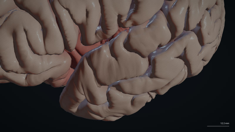
The whole brain. One brain, its own pial surface, filling the view two seconds into the dive.
MethodsBigBrain 2015's own pial surface (Amunts et al. 2013), 163,842 vertices a hemisphere, carried into MNI152 2009c through its recorded template warp. Native Metal still at 3840×2160, 2.0 s into the 18 s dive (stage 1 of 8, hd-cortex, 0–4 s), engine 3fd56e63, the 2026-10-03 powers-4k-v1 capture set with SHA256SUMS. · reference
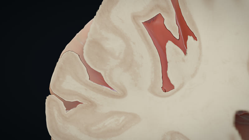
Into the cut. The camera magnifies into the left temporal lobe as the coronal cut opens between the folds.
MethodsThe pial surface fades as it magnifies, and a coronal cut sweeping back to y = −4 mm opens BigBrain 2015's histology (Amunts et al. 2013). Native Metal still at 3840×2160, 4.4 s into the 18 s dive (stage 2 of 8, cortex-histology-dissolve, 4–4.8 s), engine 3fd56e63, the 2026-10-03 powers-4k-v1 capture set with SHA256SUMS. · reference
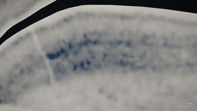
Histology at 100 µm. The cut face in Merker stain: cell-dense cortex as a dark band over pale white matter, from the sections themselves.
MethodsBigBrain 2015 at 100 µm (Amunts et al. 2013), mapped to MNI152 2009c: 64³ bricks ray-marched at 0.15 mm, the cut face in a measured Merker-stain display LUT. Native Metal still at 3840×2160, 8.8 s into the 18 s dive (stage 3 of 8, bigbrain-100um, 4.8–9.4 s), engine 3fd56e63, the 2026-10-03 powers-4k-v1 capture set with SHA256SUMS. · reference
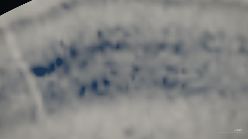
Histology to tissue. The stain at its closest, the moment before a different brain's tissue fades in at the same scale.
MethodsBigBrain's cut dissolves into H01, a separate specimen shown beside the cut at the same scale, not registered to it (Amunts et al. 2013; Shapson-Coe et al. 2024). Native Metal still at 3840×2160, 9.4 s into the 18 s dive (stage 4 of 8, histology-h01-dissolve, 9.4–10.2 s), engine 3fd56e63, the 2026-10-03 powers-4k-v1 capture set with SHA256SUMS. · reference
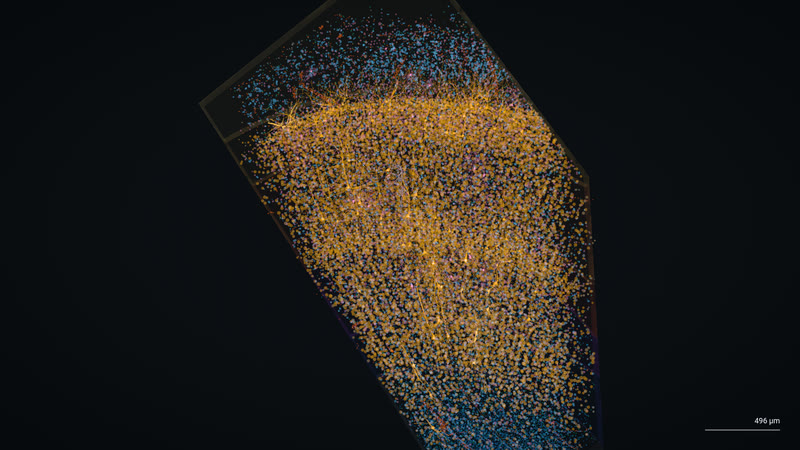
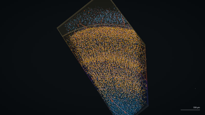
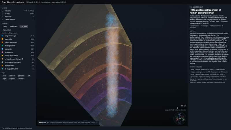
One cubic millimetre. H01's column of somata in their layers, every cell body from a cubic millimetre of human temporal cortex.
MethodsH01 (Shapson-Coe, Januszewski, Berger et al. 2024): electron microscopy of a cubic millimetre of human temporal cortex, its 57,215 cells in their cortical layers. Native Metal still at 3840×2160, 11.5 s into the 18 s dive (stage 5 of 8, h01-somata, 10.2–11.5 s), engine 3fd56e63, the 2026-10-03 powers-4k-v1 capture set with SHA256SUMS. · measured
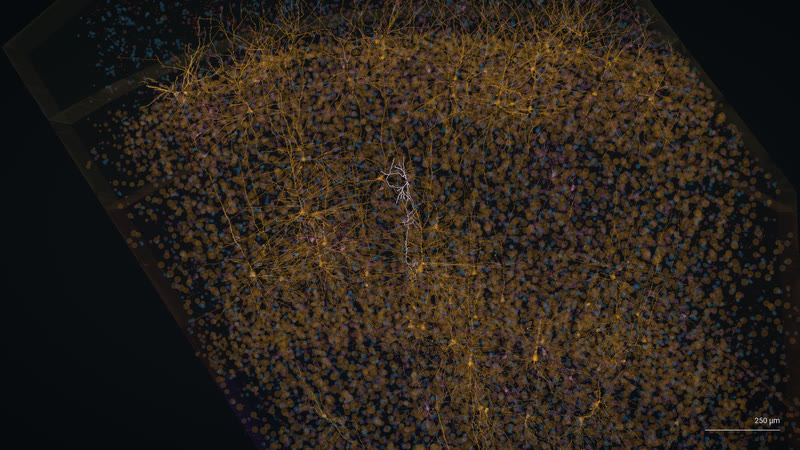
The arbors. The proofread cells' skeletons faded in across the column, each simplified to a micrometre.
MethodsThe skeletons of H01's 104 proofread cells fade in, simplified to 1 µm (Shapson-Coe et al. 2024). Native Metal still at 3840×2160, 12.3 s into the 18 s dive (stage 6 of 8, h01-arbor-reveal, 11.5–12.3 s), engine 3fd56e63, the 2026-10-03 powers-4k-v1 capture set with SHA256SUMS. · measured
Wiring. The skeletons coloured by cell type, with one cell brought forward from the rest.
MethodsH01's proofread skeletons coloured by cell type, and one of them brought forward (Shapson-Coe et al. 2024). Native Metal still at 3840×2160, 15.0 s into the 18 s dive (stage 7 of 8, h01-arbors, 12.3–15 s), engine 3fd56e63, the 2026-10-03 powers-4k-v1 capture set with SHA256SUMS. · measured
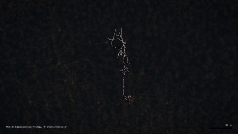
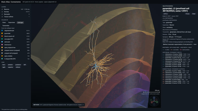
One neuron. The dive ends on a single proofread neuron with its synapses, alone in the dark of its neighbours.
MethodsOne of H01's 104 proofread neurons, with its synapses (Shapson-Coe et al. 2024). Native Metal still at 3840×2160, 18.0 s into the 18 s dive (stage 8 of 8, h01-proofread-neuron, 15–18 s), engine 3fd56e63, the 2026-10-03 powers-4k-v1 capture set with SHA256SUMS. · measured
Pulsatile flow through one real person's arteries.
A 1D network of 88 arteries everywhere and 3D CFD in the hero vessels, on IXI165's own angiogram. Then a clot lodges, the collaterals take over, and the clock starts on the tissue.
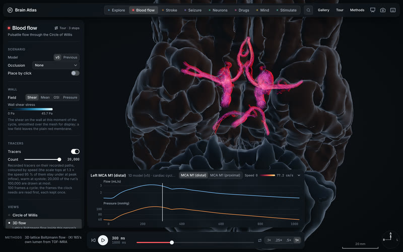
20,000 tracers ride the heartbeat. Drawn from the CFD's 100,000, through IXI165's Circle of Willis for one simulated cycle, looped three times: fast in the carotids, slow and swirling at the apices, inside a membrane that glows with wall shear stress.
Methods3D lattice-Boltzmann CFD at 0.1 mm on IXI165's TOF-MRA lumen (242,682-vertex wall), inlets from the 1D v4 model, cycle periodic to 0.3 % · Blender 5.2 Cycles, 1080p at 4.17× slower than life · simulated
A clot in the left middle cerebral artery: before and after. Distal M1 occlusion. The anterior communicating artery's flow rises from 57 to 86 mL/min as the circle compensates.
Methodsixi165-1d-v4-occl-l-m1 against the v4 baseline · wgpu engine, split capture stacked for the phone · simulated
Left carotid blocked: the circle reverses. The left A1 turns around (+134 to −111 mL/min) and 216 mL/min crosses the anterior communicating artery from right to left.
Methodsixi165-1d-v4-occl-l-ica, 15 s · Blender 5.2 Cycles · simulated
A basilar clot, and one tiny artery decides. With the traced posterior communicating artery the tissue is spared; with the main anatomy the untreated infarct reaches 153 mL by 24 hours.
Methodsixi165-1d-v4-occl-ba with the PCoA bracket · stroke-v4 tissue fate on IXI165's territories · Blender 5.2 Cycles · simulated
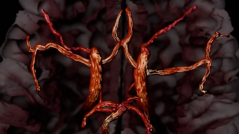
The Circle of Willis from this person's own angiogram. Centrelines and radii from the TOF-MRA where the tree overlaps the network, with the flow's particles inside.
MethodsIXI165 (IXI, 3 T TOF-MRA), TopCoW labels · run ixi165-1d-v4-baseline · Blender 5.2 Cycles, 1024 samples · measured
Arteries and veins on the living cortex. Red pial arteries and blue veins on BigBrain's surface, the fibre bundles glowing through the tissue, the arterial tree swollen by the simulated pressure of one heartbeat.
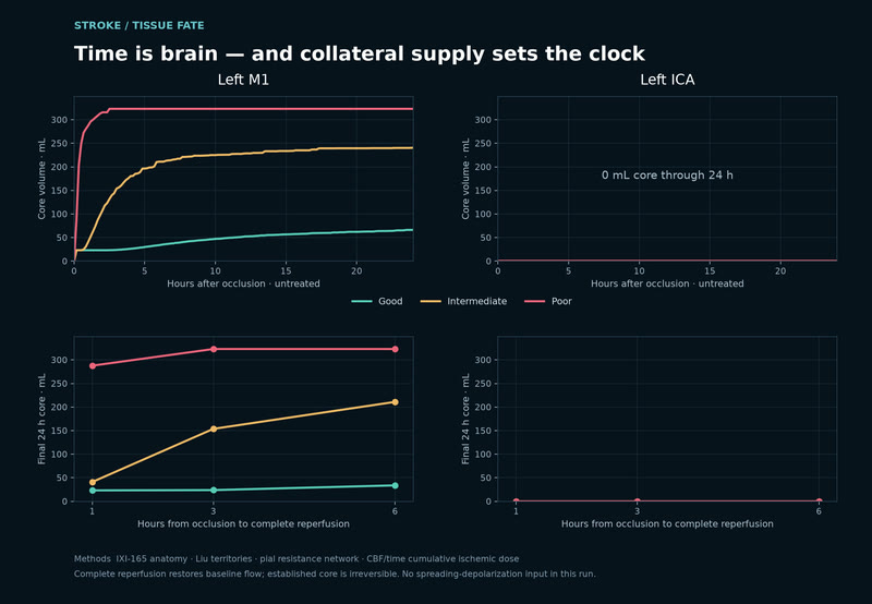
Time is brain, in millilitres. Proximal M1 clot with intermediate collaterals: reperfusion at 1 h prevents 199 mL of the 24-hour infarct, at 3 h 87 mL, at 6 h 29 mL.
MethodsTissue-fate model on IXI165's territories over the 1D v4 occlusion; untreated 24 h core 240 mL · simulated
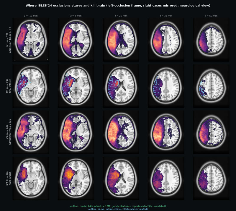
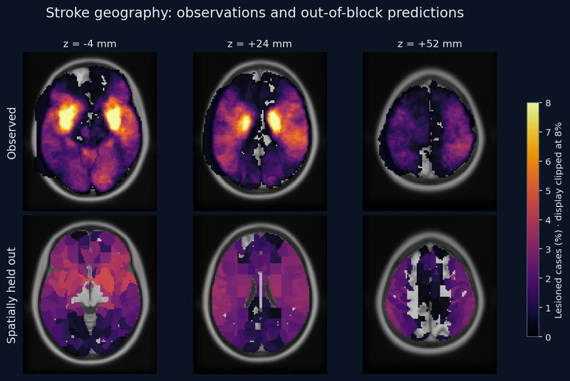
Where real strokes starve and kill brain. 149 ISLES'24 patients: at admission the Tmax > 6 s deficit fills the MCA territory; after thrombectomy the final infarct concentrates in the deep lenticulostriate bed, hit in 47 % of M1 and 57 % of ICA patients at the peak voxel.
MethodsISLES'24 training set, 149 cases registered to MNI (brain-mask Dice median 0.977); outlines are the model's 24 h infarct for a left M1 clot · measured
Blood flow, now in 3D. Twenty thousand recorded tracers ride one heartbeat through IXI165's own arteries, and the wall lights up with the shear they put on it.
Methods3D CFD of IXI165's arteries: run ixi165-cfd-v2-baseline via its display copy (header 7f8da9ec…), pulsatile D3Q19 lattice Boltzmann flow (Latt & Chopard 2006) through the TOF-MRA lumen of pack ixi-165-v0, openBF v4 Womersley inlets; 20,000 of 100,000 recorded tracers coloured by speed, and per-frame wall shear stress on a 0–45.7 Pa legend. Live app 1c351285 (embed a5d41440) at ?scene=bloodflow&view=cfd, headless capture 2880×1800. · simulated
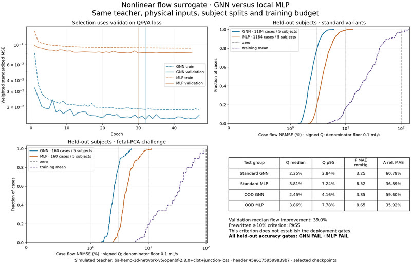
Graph messages help flow; deployment gates fail. Validation flow error falls 39%, but pressure, direction and Mac CPU timing miss the registered targets.
MethodsFailed deployment gates: GNN/MLP miss pressure, direction and the observed Mac CPU latency target; relative area, direction and reversal lose to training mean. Frozen v5 openBF r3: 10,880 simulated cases on 46 anatomies, zero/mean controls, one test evaluation per model. IXI was scored in r2; only three TUM test subjects are new. Graph messages cut validation flow NRMSE 38.98%. Shared-host timing; full methods in the 2026-10-03 r3 note. · derived
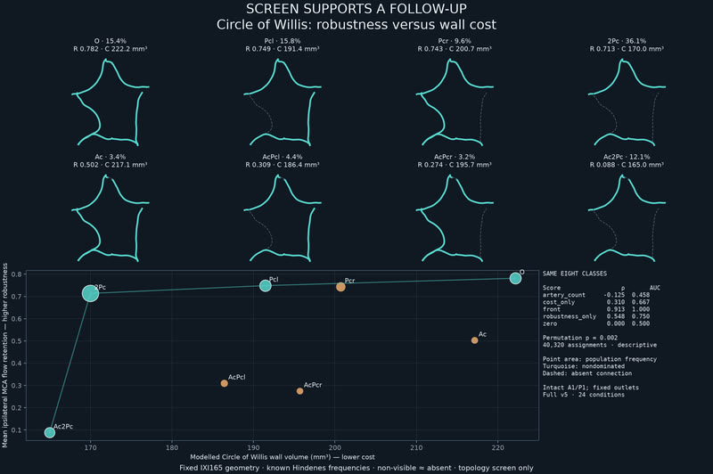
Eight circles on a robustness–cost front. Screen supports a follow-up: a fixed-anatomy screen beats four controls. Front membership is not adequate collateral perfusion.
MethodsScreen supports follow-up only. Unvalidated v5 flow on one IXI165 anatomy; eight patterns and known Hindenes frequencies (conditional N=1,434). Front rho=0.913/AUC=1.00 beats zero, cost, robustness and artery-count controls (best 0.548/0.75). Non-visible vessels count as absent. Front membership is not adequate collateral perfusion: the cheapest pattern retains 8.8% MCA flow. Whole-set gates; one score. Full methods in the research note. · simulated
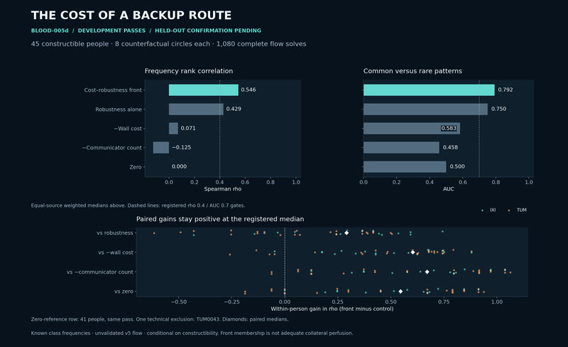
A backup route has a cost. Development passes on the median of 45 anatomies; held-out confirmation is pending.
MethodsDevelopment passes, conditional on constructible anatomy: median rho 0.546 and AUC 0.792 across 45/46 IXI/TUM people, with positive paired median gains over all four controls. The 41-person zero-reference row also passes. One pre-solver exclusion; 159 held-out cases still require confirmation. Eight counterfactual circles use unvalidated v5 flow and known class frequencies. Front membership is not adequate collateral perfusion. · derived
A spreading depolarization crosses one person's cortex. After the left M1 occlusion, a simulated depolarization wave spreads for 30 minutes until it covers a quarter of IXI165's left cortex. The engine draws it live on that person's own folds, under their own arteries.
MethodsFitzHugh-Nagumo reaction-diffusion on fsaverage, seeded by the v4 left-M1 penumbra (spreading-depolarization-ixi165-m1-v4-8563700d-t0-v1), mapped onto ixi-165-cortex-hd 0.1.0 (IXI165's FastSurfer pial surface) through its map-fsaverage; wgpu engine, native 1080p, 150× model time. · simulated
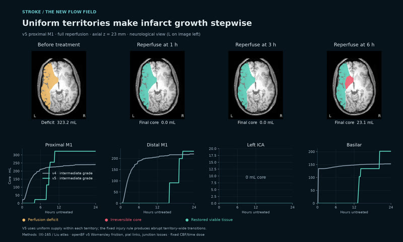
Uniform flow makes infarct growth stepwise. With the uniform v5 tissue rule, proximal-M1 core is 0, 0 and 23.1 mL after reperfusion at 1, 3 and 6 h.
MethodsIXI-165 geometry and Liu territories; openBF v5 with Womersley friction, literature pial links and total-pressure junctions, coupled to the fixed relative-CBF/time injury rule. V4 intermediate grade is compared with v5, whose three grades are identical. Neurological view: left is image left. · simulated
A seizure starts in the left mesial temporal lobe and spreads, with the scalp EEG and the depth electrodes a clinician would see. Real patients sit beside the model.
The seizure spreads across the cortex. Each parcel pulses with its own simulated discharges as the network recruits, played at 2.2× model time.
MethodsEpileptor on HCP-derived connectivity, fsaverage cortex, MNE BEM scalp EEG · wgpu engine with the bloom pass, 1080p · simulated
Beside the model: real patients. The HUP intracranial EEG dataset, 57 patients and 208 seizures with clinically marked onset channels and resections. In the app, HUP185's SEEG and HUP070's ECoG sit next to the simulation.
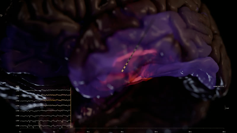
The seizure begins, under the depth electrode. 7.5 s into a simulated left mesial temporal seizure the parahippocampal cortex, the hippocampus and the amygdala light on BigBrain's own folds, seen from below and in front at 85 mm; the SEEG strip's contacts nearest the focus turn amber. The unlit hemisphere reads as dark tissue, the window is the brightest thing in the frame.
MethodsDelayed six-state Epileptor network on HCP-derived connectivity (dynamics/seizure-left-mesial-temporal-lateral-spread-distributed-v1: 82 regions, 35 s, OpenMEEG depth potentials), painted on bigbrain-cortex-hd; a modelled depth electrode and its 8 contacts' potentials in the strip · Blender 5.2 Cycles, frame 120, 1024 samples, 2560×1440 · simulated
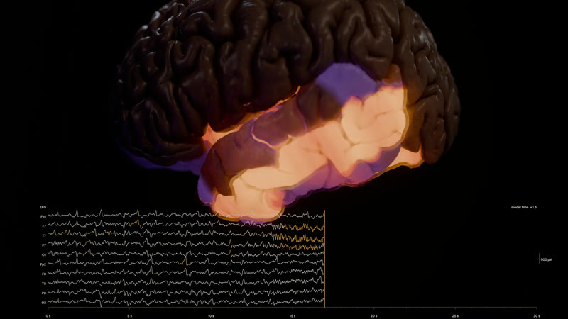
Recruitment: the temporal lobe lit at 17 s. The same seizure 17 s in, from the left: the superior and middle temporal gyri, the insula, the fusiform and the anterior cingulate are above a third of the model's activity range and the EEG's left temporal channels (F7, T7, P7) run amber. The run never crosses to the right hemisphere.
MethodsDelayed six-state Epileptor network on HCP-derived connectivity (dynamics/seizure-left-mesial-temporal-lateral-spread-distributed-v1: 82 regions, 35 s), painted on bigbrain-cortex-hd; scalp EEG from the run's MNE forward model in the strip · Blender 5.2 Cycles, frame 272, 1024 samples, 2560×1440 · simulated
One cell firing on its own reconstruction, a thousand-cell cortical circuit in health and depression, and the electron-microscopy tissue it all lives in.
A human layer 2/3 pyramidal cell fires. Two back-propagating action potentials, a basal NMDA spike, then six synaptic clusters drive an output spike.
MethodsEyal 2018 cell 0603_11 in NEURON, its own reconstruction · run eyal2018-0603-11-bap-nmda-r2 · Blender 5.2 Cycles, 2560×1440 · simulated
A living human microcircuit: 1,000 cells. Layer 2/3 at 34 °C. Firing rates PYR 1.0, SST 5.4, PV 10.1, VIP 3.4 Hz against the paper's 1.1, 5.6, 10.3 and 3.5. The EEG peaks at 11 Hz.
MethodsYao et al. 2022 human L2/3 microcircuit in NEURON + LFPy, 4.5 s with 2.5 s analysed, every compartment at 0.25 ms · run yao2022-l23net-baseline · simulated
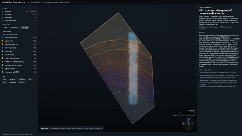
H01's column in the engine. 7,698 cell skeletons, 2,741 of them neurons, and 5.2 million synapse rows from a cubic millimetre of human temporal cortex, coloured by cell type.
MethodsH01 (Shapson-Coe et al. 2024), pack h01-patch-v0 0.2.0 · wgpu engine, 4K capture on M1 Max · measured
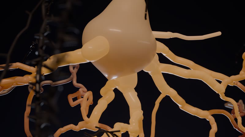
The soma under film light. A human layer 2/3 pyramidal cell glowing with its own simulated voltage, the 104 proofread H01 neighbours as ghosts behind it.
MethodsEyal 2018 cell 0603_11 in NEURON 9.0.2, synapse beads from run eyal2018-0603-11-bap-nmda · H01 proofread skeletons · Blender 5.2 Cycles, 768 samples, frame 96, 2560×1440 · simulated
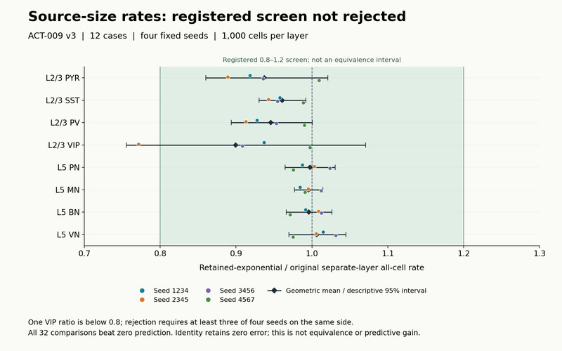
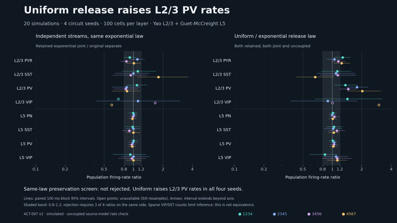
Two cortical layers pass a firing-rate screen. Giving each synapse its own random release sequence triggered no rejection compared with the original layer models run separately. Upper-layer rates ran a little low: a screen, not a match.
MethodsACT-009 v3, NOT REJECTED: Yao 2022 L2/3 + Guet-McCreight 2022 L5, NEURON/LFPy; 12 uncoupled cases, four seeds, 1,000 cells/layer. Retained-exponential/original rate ratios: VIP 0.7713; 15/16 L2/3 below 1. Band: 0.8–1.2 registered screen; intervals descriptive. All 32 comparisons beat zero; identity error is zero. No equivalence, physiological calibration or predictive superiority. L5 stimulus construction disabled. V2 failed and unscored. · derived
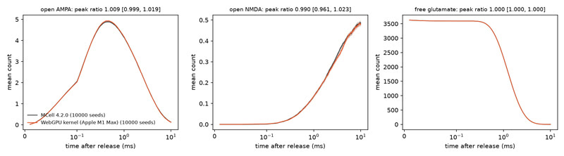
The WebGPU glutamate kernel matches MCell4 at H01 synapse e2. CELL-001 passes and is informative under the registered AO equivalence rule: open-AMPA peak 1.009 [0.9995, 1.019] and 0–10 ms integral 0.9965 [0.985, 1.009], both inside [0.95, 1.05].
MethodsWebGPU kernel ba_render::synapse (MCell's algorithm in WGSL) against MCell 4.2.0 on the same H01 e2 bundle: 97,348 faces, 80 AMPA and 30 NMDA sites, 3,640 glutamate, dt 0.5 µs; 10,000 seeds per arm, 90 % bootstrap intervals, registered before any scoring run on e2. PHARMA-002's box fails the same rule (peak 1.52), so the test is informative. Every score output reproduced byte for byte from the retained files. H01: Shapson-Coe et al. 2024. · simulated
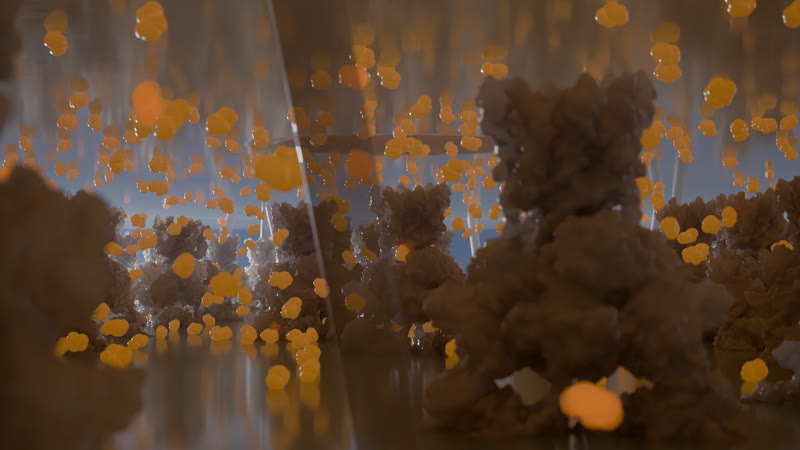
Glutamate in H01 synapse e2, live on the GPU. One vesicle's 3,640 glutamate cross the cleft and bind AMPA and NMDA receptors, a Monte Carlo simulation run live in the browser and shown 10,000 times slower than real time.
MethodsMonte Carlo simulation, live on the GPU (MCell's algorithm in WGSL): 3,640 glutamate from one docked vesicle diffuse in 0.5 µs steps and bind 80 AMPA and 30 NMDA sites by Singh et al. 2011's kinetics. Measured (H01, Shapson-Coe et al. 2024): e2's membranes, PSD and docked vesicles. Derived: the 20 nm cleft. Placed, not observed: the receptor sites. CELL-001 checked the kernel against MCell 4.2.0 on the same mesh, sites and rates, within ±5 %. · simulated
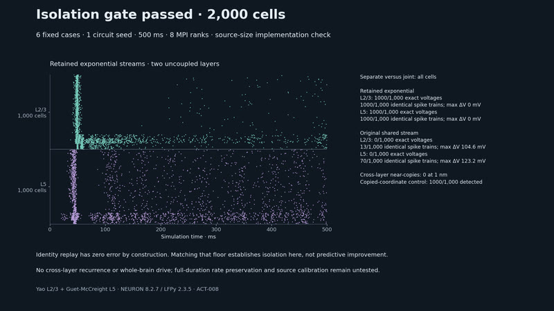
Two layers, isolated streams. Isolation gate passed: 2,000 cells match separate-layer traces for one seed over 500 ms; identity replay also passes, and the L2/3 startup burst dominates.
MethodsImplementation isolation, one seed, 500 ms, identity floor: ACT-008 uses Yao L2/3 and Guet-McCreight L5 in NEURON/LFPy, 1,000 cells per layer and eight MPI ranks. Retained exponential streams reproduce separate-layer traces and spikes exactly; the shared stream fails. Only 272 L2/3 and 685 L5 cells spike in [100,500) ms; 72% of L2/3 spikes occur before 100 ms. Full-duration rates, cross-layer coupling and whole-brain drive remain untested. · simulated
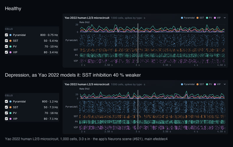
A human microcircuit fires, healthy and in depression. Yao 2022's human L2/3 circuit, 1,000 cells over 2.5 s, in the app's Neurons scene. Healthy, its pyramidal cells fire at 0.75 Hz, SST 5.4, PV 10, VIP 3.4. With SST inhibition 40 % weaker, the paper's depression condition, every type speeds up: pyramidal 1.2 Hz, SST 7.3, PV 16, VIP 7.1, 7,472 spikes against 4,618.
MethodsYao et al. 2022 human L2/3 cortical microcircuit (Cell Reports 38, 110232) in NEURON 8.2.7 at 34 °C: 800 pyramidal, 50 SST, 70 PV and 80 VIP cells on replicated Allen human morphologies; runs cells/yao2022-l23net-baseline and cells/yao2022-l23net-depression, depression the paper's MDD condition with SST synaptic and tonic inhibition reduced by 40 %; rates and rasters are each run's own somatic spikes over the recorded 2.0–4.5 s (metrics.json: pyramidal 0.7525 against 1.2005 Hz; the paper reports 0.77 and 1.2); shown by the app's Neurons Model → Microcircuit at 3.0 s, the circuit not yet drawn in the 3D view · simulated
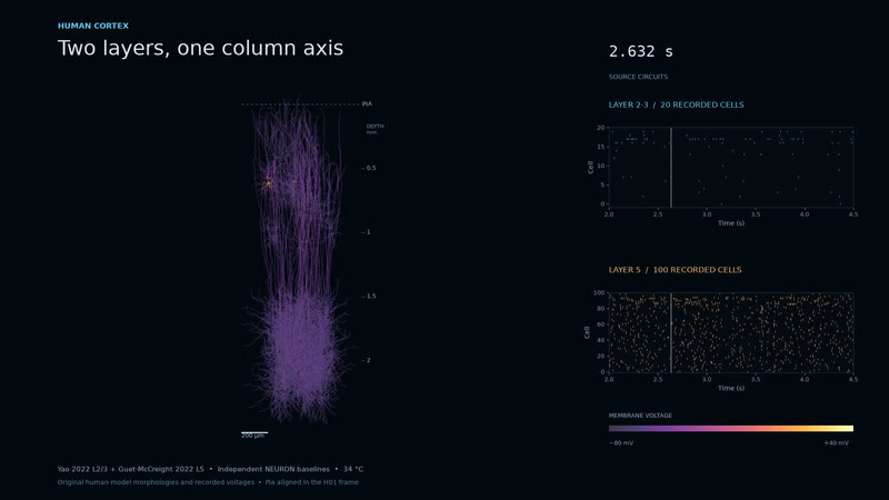
Two layers, one column axis. Yao 2022's human layer 2/3 circuit (20 recorded cells) sits over Guet-McCreight 2022's layer 5 (100 cells) on a shared pia axis, as voltage-coloured morphologies with their rasters. Two independent NEURON baselines aligned in the H01 frame, not yet a coupled column: the coupled run is the next bet.
MethodsRuns yao2022-l23net-baseline-closeup-r2 and guetmccreight2022-l5net-test100-r2 (NEURON), frames at 2.632 s, placed by rigid translation in the h01-patch-v0 0.2.1 column frame · derived
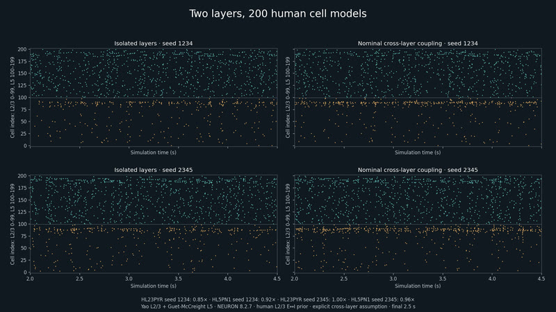
Two layers, 200 human cell models. Nominal coupling raises L2/3 PV spike counts 1.73× and 1.71× in two simulated seeds.
MethodsYao L2/3 and Guet-McCreight L5, 100 cells each in NEURON 8.2.7; source OU input, human-informed within-L2/3 E↔I probabilities, assumed Potjans–Diesmann cross-layer wiring, retained uniform synapse RNG. Raster: final 2.5 s of 4.5 s runs, human-isolated versus human-coupled, seeds 1234 and 2345 (ACT-002). · simulated
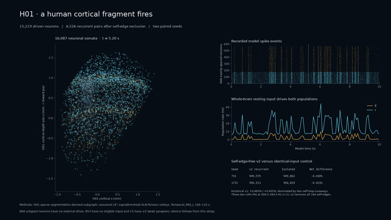
Human cells, whole-brain input. 15,219 driven neurons fire in 10-second simulations; removing 104 self-edges eliminates the runaway cells.
MethodsH01 soma locations and sparse segmentation-derived subgraph; assumed exponential-current LIF with suprathreshold Poisson volleys from ALN Temporal_Mid_L, 100–110 s. Post-hoc self-edge-free v2, 6,526 recurrent pairs, two identical-input controls. Untyped neurons have no external drive; 853 have no eligible input and 15 have at most five weak synapses. Pack h01-patch-v0 0.3.0, default h01-lif-resting-recurrent-seed731-v2. · simulated
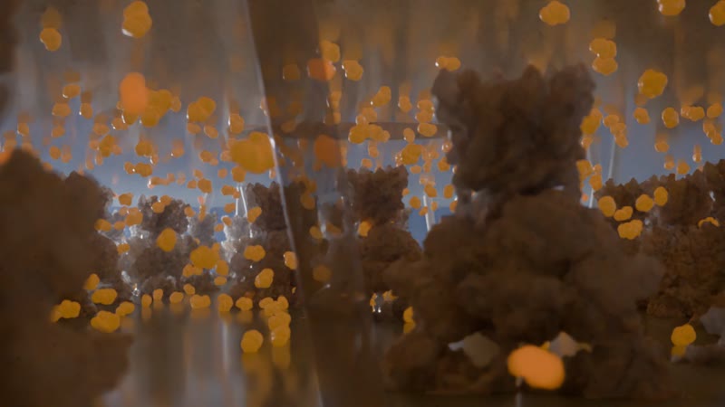
Inside the synaptic cleft. Glutamate crossing the 20 nm cleft 100 microseconds after release: a burst of 1,500 molecules from one fused vesicle, binding AMPA and NMDA receptors on the spine. Frame 430 of the neurons sequence, 2560×1440.
MethodsMolecule positions from the Smoldyn run synapse-glutamate-nbqx-zero-drug (1,500 glutamate every 20 µs for 43 ms in a 300 nm × 20 nm cleft, 80 AMPA + 20 NMDA sites with kinetic states), drawn where the run puts them; receptors are PDB 3KG2 (GluA2) and 7SAA (GluN1/GluN2B) as Molecular Nodes surfaces at the run's sites; the bouton, vesicle, pore, spine and membranes are modelled from literature dimensions. Cycles, 768 samples, 85 mm at f/0.35, the neurons scene at 1 nm = 1 m. · simulated
Receptors bind by real densities and affinities across the cortex, and at the synapse, molecules meet their receptors one at a time.
1,500 glutamate molecules, with and without the blocker. A 43 ms synaptic event as particles: NBQX competes for the AMPA receptors and cuts their integrated open time by 87 %.
MethodsSmoldyn particle simulation, Jonas AMPA and Erreger NMDA kinetics, 80 AMPA and 20 NMDA receptors on a 300 × 300 nm face · 12-seed ensemble: 87.45 % lower AMPA open time · run synapse-glutamate-nbqx · simulated
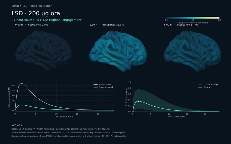
LSD, 200 µg, over 24 hours. Plasma, free brain concentration and 5-HT2A occupancy, peaking at 35 % at 1.6 h, painted on the cortex by receptor density.
MethodsDolder 2017 oral PK, frozen Ki binding, Beliveau 2017 5-HT2A PET map on fsLR · run lsd-200ug-cortex-v1 · simulated
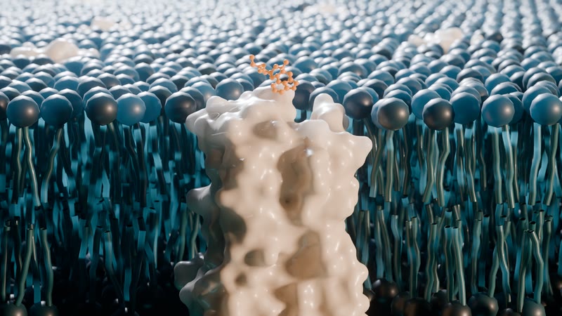
LSD seated in its receptor. The 5-HT2A receptor cut open in its lipid bilayer, the drug's 24 heavy atoms in the crystal pose as the receptor shifts to its drug-bound state; at the peak, 45 of 127 receptors carry it.
MethodsPDB 6WGT (LSD-bound 5-HT2A) morphing to 9WPQ, Molecular Nodes; occupancy from the PK course, 96 min after 200 µg · Blender 5.2 Cycles, frame 241, 512 samples, 2560×1440 · reference
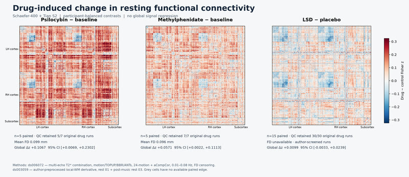
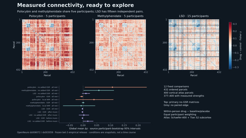
Measured, not modelled: how three drugs change connectivity. Global shift in Fisher z: psilocybin +0.107, methylphenidate +0.057, LSD +0.010. With global-signal regression, psilocybin and LSD turn negative and methylphenidate stays just above zero. The receptor-informed model is the next step.
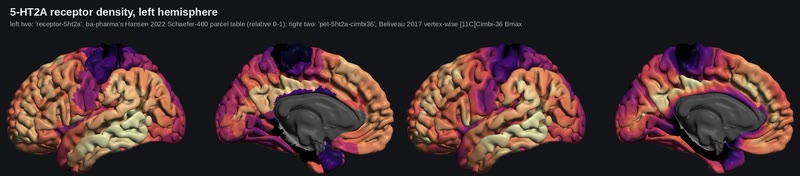
5-HT2A density on the HD cortex. The receptor map the drug scenes paint with: the Schaefer-400 parcel table on the left, the vertex-wise PET map on the right.
MethodsHansen 2022 parcel tables and Beliveau 2017 vertex-wise 5-HT2A PET, on the cortex-hd pack · measured
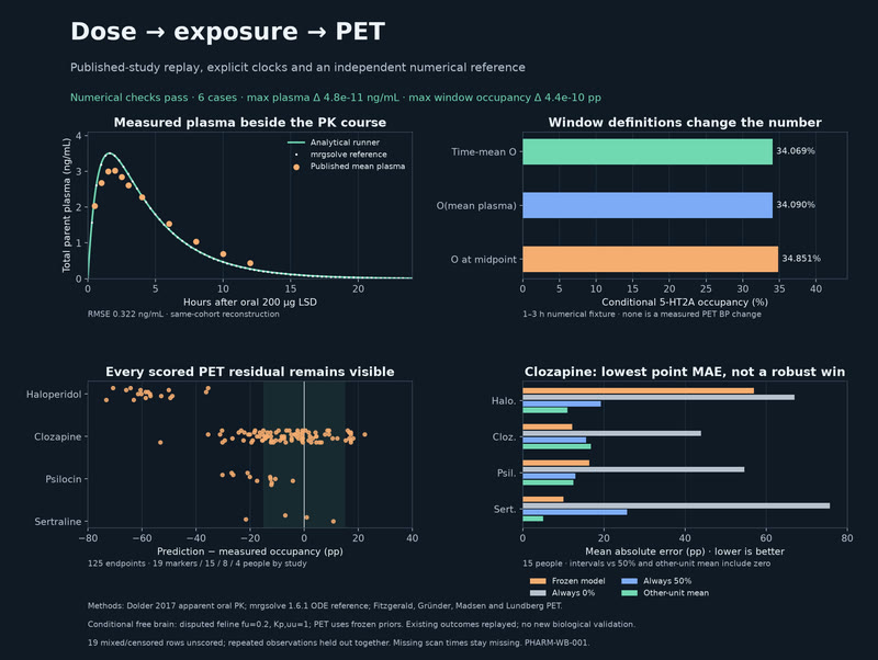
Dose, exposure and PET: reproduce the evidence. 10,125 samples agree with mrgsolve; four PET studies keep their measured errors and trivial baselines visible.
MethodsNumerical pass; conditional pharmacology: PHARM-WB-001/workbench-replay-v2, analytic PK vs mrgsolve 1.6.1; 6 cases, 40 windows. Four known PET studies replayed exactly; clozapine's model has the lowest point MAE in its study of 15 people, not a robust win. Human fu/Kp,uu unknown; disputed feline fu=0.2 used for LSD. Missing scan times stay null; 19 mixed/censored rows unscored. No tracer model or new biological validation. See 2026-10-04-pharma-workbench-replay. · derived
Measured changes in drug FC. Preprocessing-dependent: 12 paired drug-FC contrasts from 5 shared psilocybin/methylphenidate participants and 15 LSD participants retain 377–400 cortical parcels.
MethodsPaired resting fMRI contrasts from OpenNeuro ds006072 (psilocybin and methylphenidate, 5 shared participants) and ds003059 (LSD, 15 participants). Schaefer-400 plus Tian S2; drug minus baseline/placebo Fisher-z FC. Twelve fixed preprocessing/rest comparisons; LSD retains its authors' preprocessing and nogsr means no additional GSR. Cortex is parcel expansion on fsLR32k. Missing cortical parcels show as bare tissue. · derived
TMS, tDCS, deep-brain and focused-ultrasound stimulation as the fields they make in real anatomy, and how the connected brain responds.
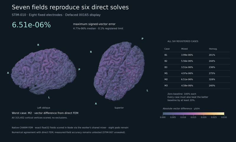
Eight electrodes, seven fields. Mixing seven precomputed lead fields with the browser tool's own mixer reproduced six direct FEM solves to within 0.00000651 % vector error; the registered limit was 0.1 %.
MethodsSTIM-010: numerical PASS. SimNIBS 4.6 FEM on native IXI165 CHARM (only the display scalp is defaced), all eight pads present in every scored solve. Float32 fields scored in Node with the browser tool's own mixer. Six cases: area-weighted relative vector error ≤0.1% and ≤0.8× the better trivial baseline. CHARM anisotropic-image and invalid-dot warnings retained. Tissue labels unvalidated; no measured-field comparison; browser latency untested. · derived
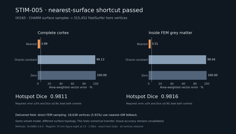
TMS on the hero cortex. IXI165's C3 field sampled directly onto the 315,452 vertices of the app's hero cortex.
MethodsSTIM-005 passed: nearest transfer 3.99% vector error, 0.9811 hotspot Dice; zero/constant 100%/99.13%. Delivered field uses direct SimNIBS 4.6 GM sampling on IXI165 FastSurfer hero vertices, C3, 1 MA/s. Same FEM, no independent tissue truth. CHARM anisotropic-input and final-label dot warnings retained. All 5.91% nearest-GM fallbacks retained (max 10.24 mm). Static pose, no IXI drag basis. Hero scalp is context, not the FEM scalp. · simulated
One field, two cortical meshes. The nearest-sample shortcut passes its registered transfer test: 3.99% vector error and 0.9811 hotspot Dice.
MethodsSTIM-005 passed: nearest transfer 3.99% vector error, 0.9811 hotspot Dice; zero/constant 100%/99.13%. Delivered field uses direct SimNIBS 4.6 GM sampling on IXI165 FastSurfer hero vertices, C3, 1 MA/s. Same FEM, no independent tissue truth. CHARM anisotropic-input and final-label dot warnings retained. All 5.91% nearest-GM fallbacks retained (max 10.24 mm). Static pose, no IXI drag basis. Hero scalp is context, not the FEM scalp. · derived
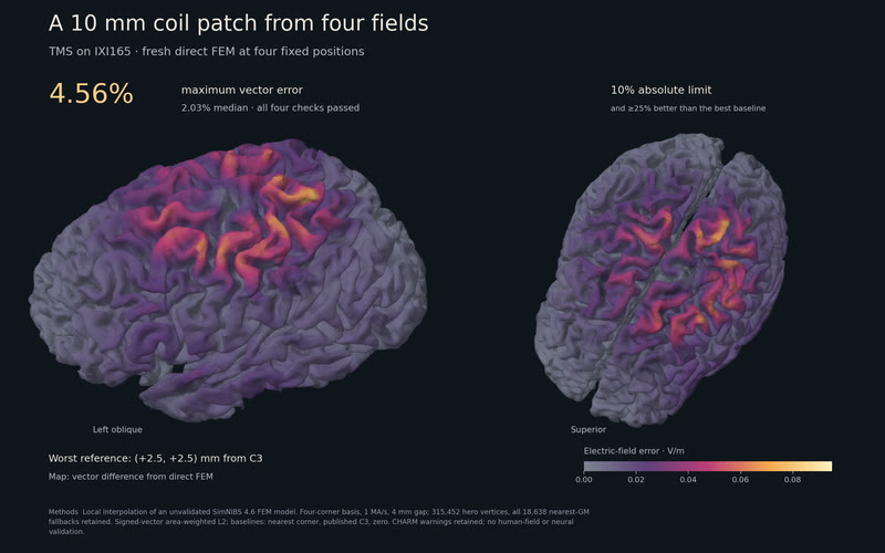
A 10 mm coil patch from four fields. Four fields matched four fresh FEM positions within 4.56% vector error; every check beat the strongest baseline.
MethodsSTIM-008 passed: the four-corner drag basis on IXI165 matched four fresh FEM positions at 2.03% median and 4.56% maximum vector error; every reference ≤10% and ≤0.75× the best baseline (nearest corner, published C3, zero). Corners (±5, ±5) mm, references (±2.5, ±2.5) mm, 1 MA/s, 4 mm gap; unvalidated SimNIBS 4.6 FEM model. Map: worst reference, (+2.5, +2.5) mm, V/m. Local numerical comparison only; no human-field or neural validation. · derived
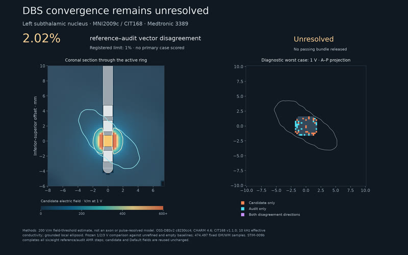
DBS convergence remains unresolved. Completing all six/eight refinements still leaves 2.02% field-vector disagreement. The registered limit was 1%.
MethodsSTIM-009b: all 6/8 reference/audit AMR steps (7/9 solutions). Signed-vector L2 2.0189% (limit 1%); VTA Jaccard 0.688/1.502/1.396% at 1/2/3 V (limit 2%). No primary case scored. Candidate/Default unchanged; 474,497 GM/WM samples. Diagnostic candidate errors 2.938/2.807/2.293%, median 2.807%, max 2.938% at 1 V. MNI152NLin2009cAsym, CHARM 4.6, CIT168 v1.1.0; OSS-DBSv2 c8230cc4, NGSolve 6.2.2601, Medtronic 3389 contact 2, 10 kHz real conductivity, grounded local ellipsoid. 200 V/m field-threshold estimate, not an axon or pulse-resolved model. Seal d266f1bf, 62 synthetic controls and 17/17 input gate; earlier path-only denial retained. Unresolved: no bundle, looser gate or retry. · derived
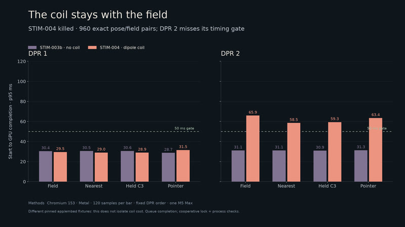
The coil stays with its field. Every pose matches its field, but full-resolution updates miss the 50 ms timing gate.
MethodsSTIM-004 killed: DPR 2 field/pointer p95 65.86/63.42 ms exceeds 50 ms; nearest allowance also fails. All 960 submitted pose/field pairs match. Ernie, 2,712 dipole glyphs; app 3e52af44, embed 6cd3e972, Chromium 153/Metal. Lock and boundary checks clean; headed apps and the compositor and short intervening jobs uncontrolled. One M5 Max, fixed DPR order, local Vite. Queue completion only; fixture comparison does not isolate coil cost. · derived
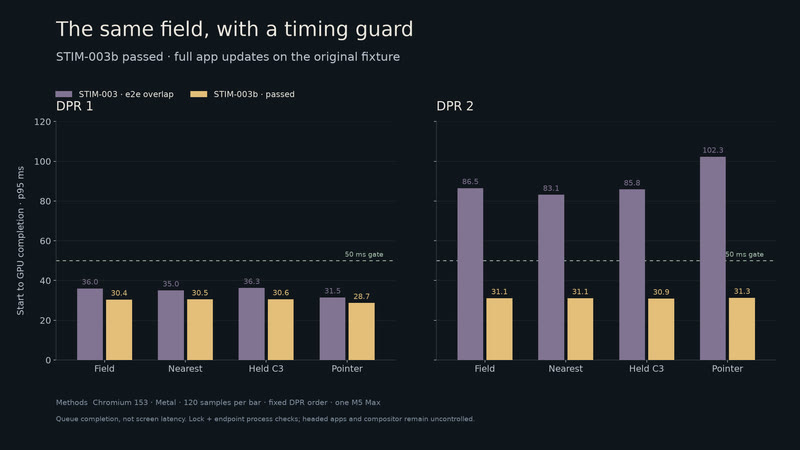
The field meets its timing gate. A guarded repeat brings full-resolution field and pointer updates to 31.10 and 31.31 ms at p95.
MethodsSTIM-003b passed: Ernie, Chromium 153 / Metal, 120 samples per condition. Lock and before/after process checks found no other headless browser at the boundaries. DPR 2 field/pointer p95 31.10/31.31 ms. Queue completion only; headed apps and the compositor and short uncooperative jobs remain uncontrolled. Fixed DPR order, one M5 Max, original embed without coil mesh. STIM-003 stays killed with its e2e overlap annotated. · derived
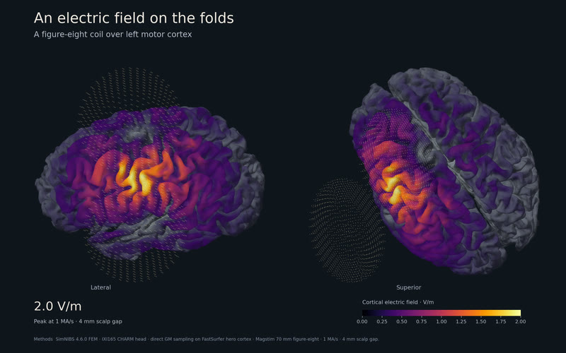
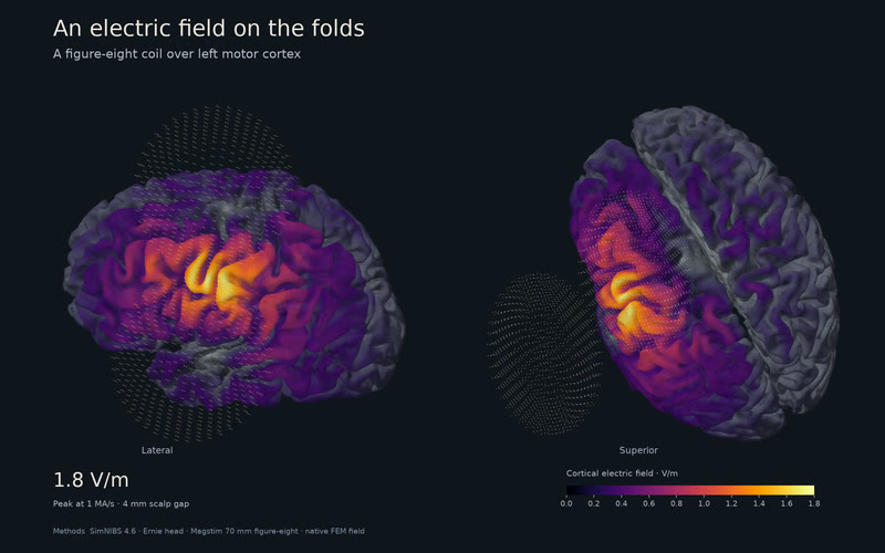
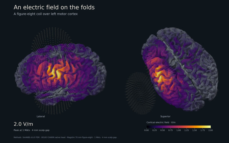
An electric field on the folds. A figure-eight coil drives a real head model: 308,791 cortical vertices, 1.77 V/m peak at 1 MA/s.
MethodsSimNIBS 4.6.0 FEM, Ernie v4.1 native head, Magstim 70 mm figure-eight at C3 with handle toward CP3, 4 mm scalp gap. Signed E mapped to the native middle surface; 6.07% of vertices use nearest-gray-matter fallback. Run ernie-tms-c3-v1. · simulated
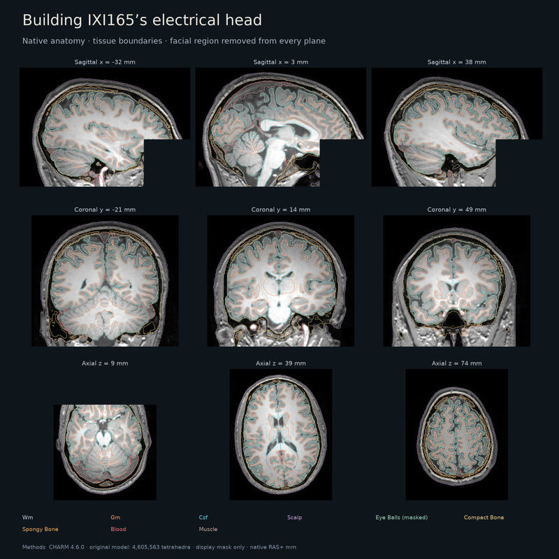
IXI165, from MRI to an electrical head. Nine tissue classes and 4.61 million tetrahedra, reconstructed from the original T1 and T2 scans.
MethodsUnvalidated: automated tissue accuracy has no independent reference. SimNIBS CHARM 4.6.0, CPU TopoFit, original IXI165-HH-1589 T1/T2. Nine native RAS+ planes; facial MRI and tissue contours are blanked before sampling with a conservative mask around both eyes, extended anteriorly and inferiorly. The original model is unchanged: nine tissue classes, 824,586 nodes, 4,605,563 tetrahedra, zero inverted or zero-volume elements. CHARM anisotropic-image and final-label dot warnings remain. Display revision only; no FEM solve or rescore. · derived
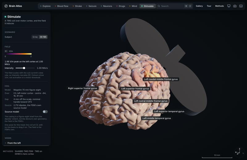
TMS on the IXI165 cortex. 1.99 V/m peak at 1 MA/s, from a finite-element solve on the subject’s own head.
MethodsUnvalidated tissue accuracy: no independent reference. CHARM warnings retained: "Anisotropic image, meshing may contain extra artifacts" and "RuntimeWarning: invalid value encountered in dot" during final labels. SimNIBS 4.6.0 FEM, original IXI165 T1/T2 (CC BY-SA 3.0), Magstim 70 mm figure-eight at C3, 1 MA/s. All 491,524 vertices retained, including 2.11% nearest-GM fallbacks (max 10.32 mm). Run ixi165-tms-c3-v2. · simulated
The cortex folding, ageing, and disease spreading along the network: measured atlases and simulated years.
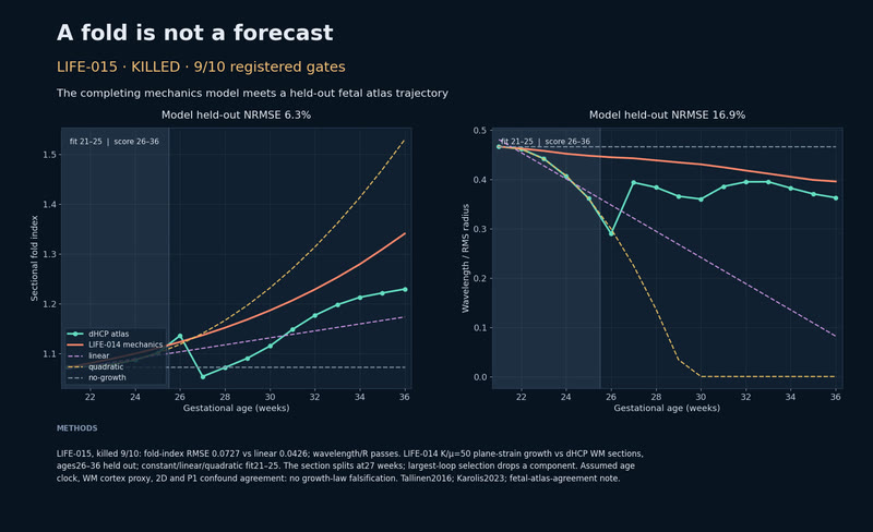
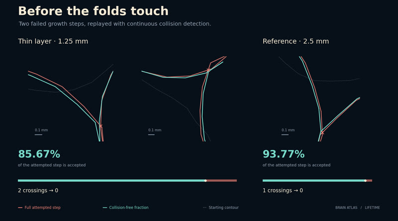
One shape. Four growing layers. Four thicknesses fold from one fetal outline; the numerical gate stops the wavelength test.
MethodsLIFE-020, NOT SCORED: refinement 2/8 over 10 % (h2/g2.5 10.45 %, h3/g2 10.52 %). 2D neo-Hookean/IPC Newton, K/μ=50, 21-week dHCP WM. LIFE-020b reran the 2 mm case with every triangle split into four: at g = 2 its fold wavelength moved 21 % (limit 10 %), GI and energy 3 % and 2 %, so the 2 mm wavelength is not mesh-converged here. The cause (P1, material sampling, contact or branch choice) is not isolated. Tallinen 2014/2016; LIFE-020/b notes. · derived
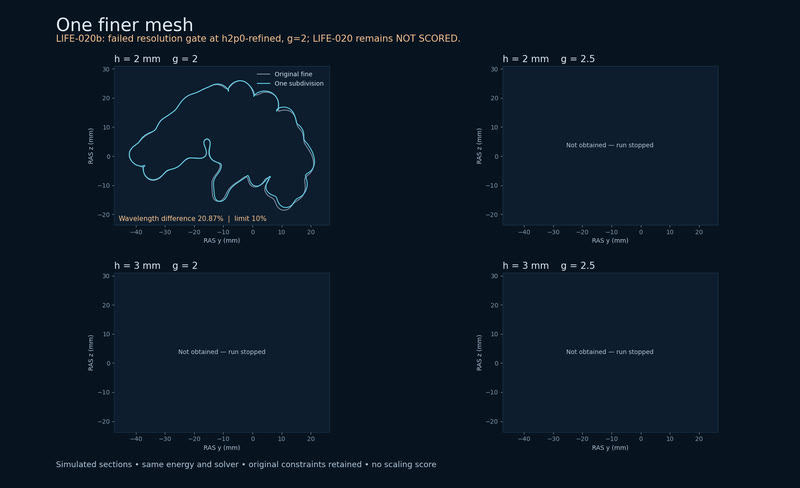
One finer mesh. The first finer-mesh comparison misses the wavelength bound. The run stops, leaving three pairs unrun.
MethodsLIFE-020b, failed refinement: h=2 mm, g=2 wavelength changes 20.87% against the frozen 10% bound; three pairs were not run. One uniform subdivision; 2D neo-Hookean/IPC Newton, K/μ=50, on 21-week dHCP WM. All 17 obtained loads pass mechanics; GI/energy differ 3.33/1.88%. P1, material sampling, contact discretization and branch choice limit inference. LIFE-020 remains NOT SCORED; no scaling prediction scored. LIFE-020b note. · derived
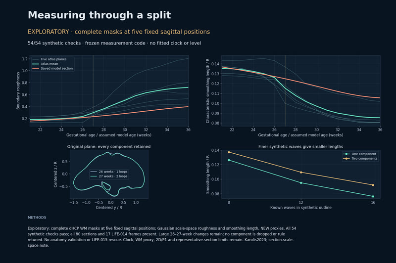
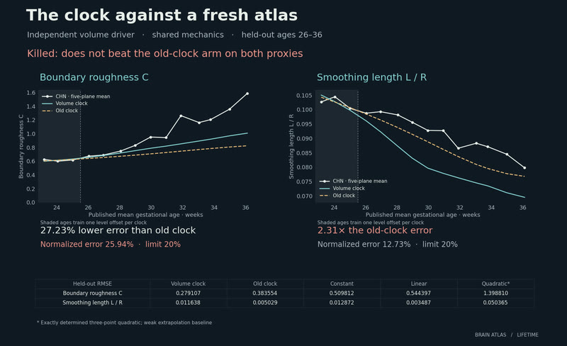
Measuring through a split. All components stay in the measurement. Five fixed sections reveal where the growing layer and the atlas differ.
MethodsExploratory: complete dHCP WM masks at five fixed sagittal positions; Gaussian scale-space roughness and smoothing length, NEW proxies. All 54 synthetic checks pass; all 80 sections and 17 LIFE-014 frames present. Large 26–27-week changes remain; no component is dropped or rule retuned. No anatomy validation or LIFE-015 rescue. Clock, WM proxy, 2D/P1 and representative-section limits remain. Karolis2023; section-scale-space note. · derived
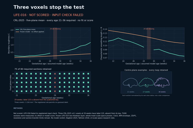
Not scored: three undefined voxels. The fresh atlas failed the registered input check. LIFE-016 supplies no model result either way.
MethodsNot scored: LIFE-016 failed its registered input check. Three CRL-2025 v4.1 voxels at 29 weeks have label 125, absent from its key. 75/80 sections were measured; no offset or model score. Frozen LIFE-014 neo-Hookean layer; whole-mask scale-space proxies. Clock, WM-envelope, 2D/P1, resolution and section-transfer limits remain. No model verdict. Bagheri 2025; Tallinen 2016; crl-scale-space research note. · derived
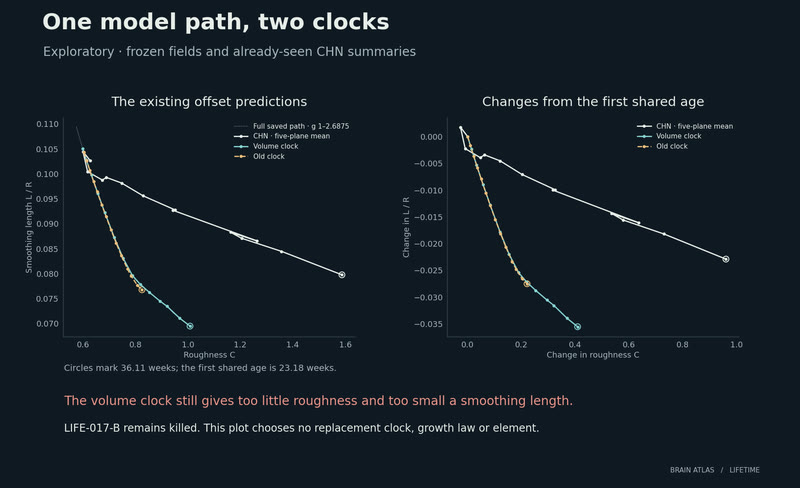
One model path, two clocks. The faster clock raises roughness but pushes smoothing length farther below the atlas.
MethodsExploratory figure: frozen LIFE-017-A model states and already-seen CHN five-plane summaries, with LIFE-017-B’s existing clock predictions and training offsets. No new solve, fit, score or gate. The second panel subtracts each series at age23.18. LIFE-017-B remains killed; the volume clock raises roughness but pushes smoothing length farther below the atlas. Constant thickness, absent core growth, WM proxy and 2D/P1 confound interpretation. Ren2022; Xu atlas. · derived
Around the time of birth. Population-average white-matter surfaces at 37, 40 and 44 weeks, seen from the left and above.
MethodsDerived reference: dHCP white-matter atlases at 37, 40 and 44 weeks postmenstrual age; display assembly: mirror-symmetry pose; hemispheres placed with a 2 mm display clearance by convention; not measured anatomy or author coordinates. Mirror mean/p95: 0.694/1.999, 0.618/1.703 mm. Volume fit failed the 1/3 mm limits. One rigid transform per side preserves v2 motion. Linear interpolation is derived. Bozek 2018; Williams 2021; neonatal-common-frame note. · derived
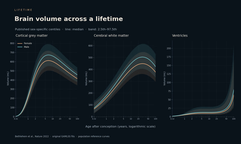
Brain volume across a lifetime. Published normative-model centiles for grey matter, white matter and the ventricles trace prenatal growth, maturation and aging.
MethodsNormative-model outputs: Bethlehem 2022 original BrainChart GMV, WMV and ventricular-volume 2.5/50/97.5 centiles, by sex. Bands show population variation. Native age is post-conception days; no neonatal-PMA conversion is assumed. Published reference curves average processing effects; the absent-fs_version contrast warnings are retained. No IXI calibration or template-volume matching. · derived
Cortical thickness across a lifetime. From age 2 to 100, regional thickness changes across one fixed reference brain. The clip follows the female reference; the export carries both female and male medians.
MethodsNormative-model reference: Rutherford 2022 Destrieux thickness, ages 2–100, female/male. Author PCNtoolkit 0.20 spline/inverse warp; site indicators zero. 148 exact parcels on fixed fsaverage, no DKT transfer or geometric aging. Source-curve replay error 0.0034 mm; no new-cohort validation. Native mesh QC retains four zero-area faces and 164k vertices/hemisphere. doi:10.7554/eLife.72904; Rutherford tooling note. · reference
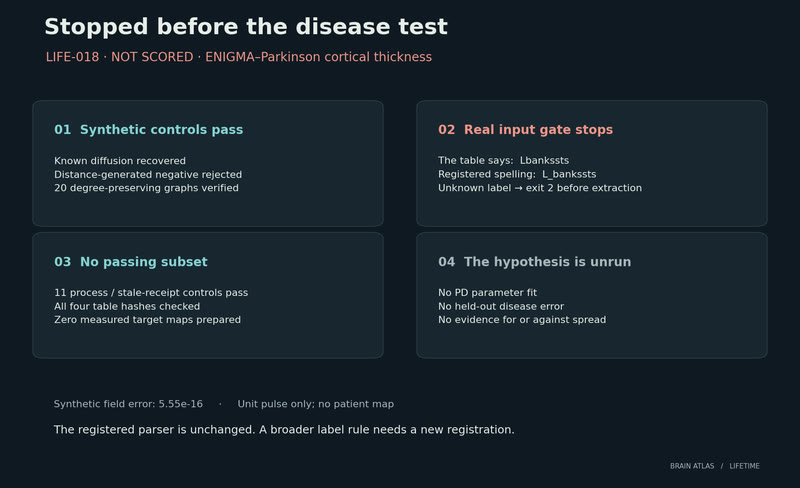
Stopped before the disease test. The label gate stopped before any Parkinson map was measured. The hypothesis remains unrun.
MethodsLIFE-018, not scored: ENIGMA-PD stage tables failed the registered DK label rule (Lbankssts versus L_bankssts). The separate input gate exited before any map extraction, fit or disease score. Synthetic structural/distance controls and 20 degree-preserving rewires passed; 11 halt/receipt checks passed. No evidence for or against disease spread. Cross-sectional stages are not elapsed time. Laansma2021; ENIGMA b08974b5; pd-diffusion research note. · derived
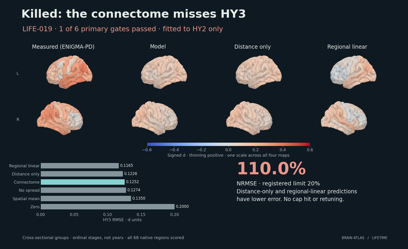
The connectome misses the next stage. Distance-only and regional-linear predictions beat the connectome; normalized error is 110%, against a 20% limit.
MethodsLIFE-019, killed: 1/6 primary; NRMSE 110% versus 20%. Distance-only 0.1226 d and regional-linear 0.1165 beat the model’s 0.1252. ENIGMA-PD HY1 seed / HY2 fit / HY3 (Hoehn and Yahr stage 3) test; no retuning. This fixed ordinal cross-sectional atrophy-pattern extrapolator fails; it does not reject protein spread. Shared controls/cohort, young-adult connectome and cortical-only coverage remain. 68 DK scored; 62 DKT displayed, 6 no data. Laansma2021; ENIGMA b08974b5. · derived
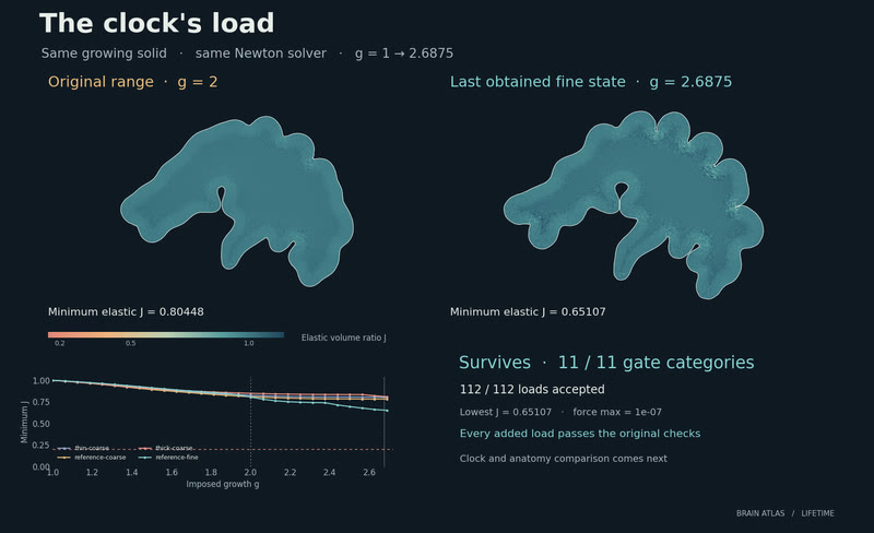
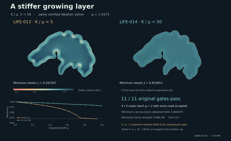
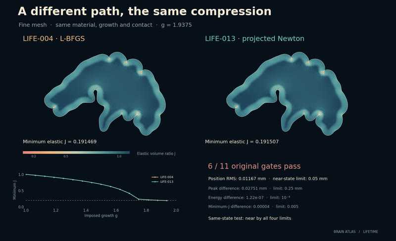
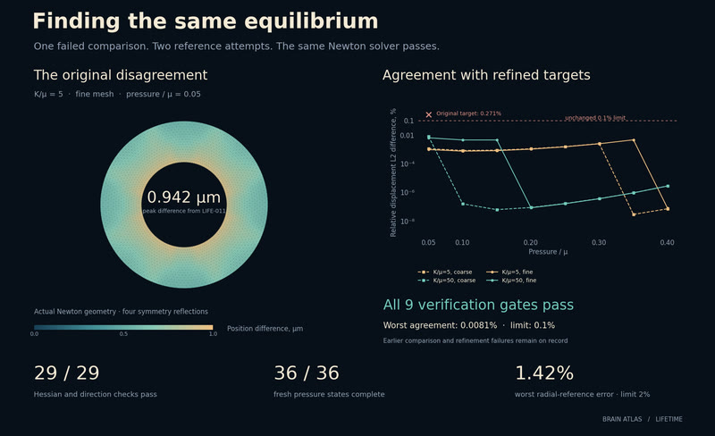
A different path, the same compression. Verified Newton returns to a nearby compressed state; the original minimum-J gate still fails.
MethodsLIFE-013, killed: verified projected Newton on the unchanged plane-strain neo-Hookean/IPC model and 21-week dHCP WM contour, assigned layers. Fine g=1.9375 reaches J=0.191507<0.2 at force convergence. All four registered near-state checks pass versus LIFE-004 (RMS 0.0117 mm); 6/11 gates pass. The selected equilibrium remains compressed; no uniqueness or anatomical claim. LIFE-004 stays killed. Tallinen 2016; IPC 1.6.0; fetal-newton note. · derived
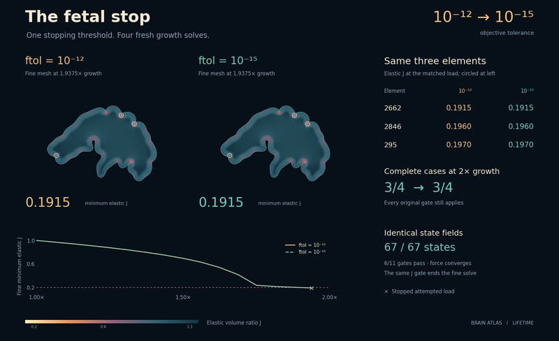
The fetal stop. A thousandfold tighter objective threshold reproduces the same compressed fetal-derived state.
MethodsLIFE-012, killed: ftol 1e-12→1e-15 reproduces every array in 67 LIFE-004 states. Fine stops at g=1.9375, J=0.19147<0.2 despite force convergence; 6/11 gates pass. Neo-Hookean plane-strain growth, K/μ=5, IPC 1.6.0, 21-week dHCP WM contour; assigned layers. This rules out the ftol-only explanation, not a specific formulation defect. LIFE-004 stays killed. Tallinen 2016; atlas doi:10.12751/g-node.qj5hs7; IPC doi:10.1145/3386569.3392425. · derived
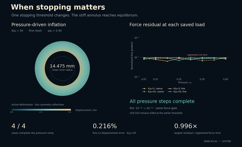
When stopping matters. A tighter objective threshold lets both stiff meshes meet the unchanged force gate.
MethodsLIFE-011, survives: changing only L-BFGS-B ftol from 1e-12 to 1e-15 completes all 36 loads and passes 15/15 gates. Plane-strain neo-Hookean annulus, K/μ=5/50; frozen radial reference. K50 fine L2 is 0.216%, beating linear by 99.52% and zero by 99.78%. LIFE-010 remains killed: its objective-change stop preceded force convergence. This does not diagnose fetal stops. SciPy 1.15.3; annulus-stopping research note. · derived
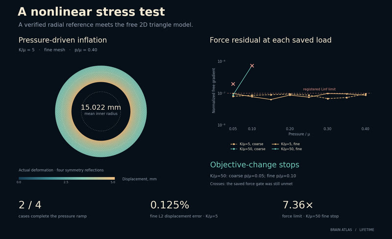
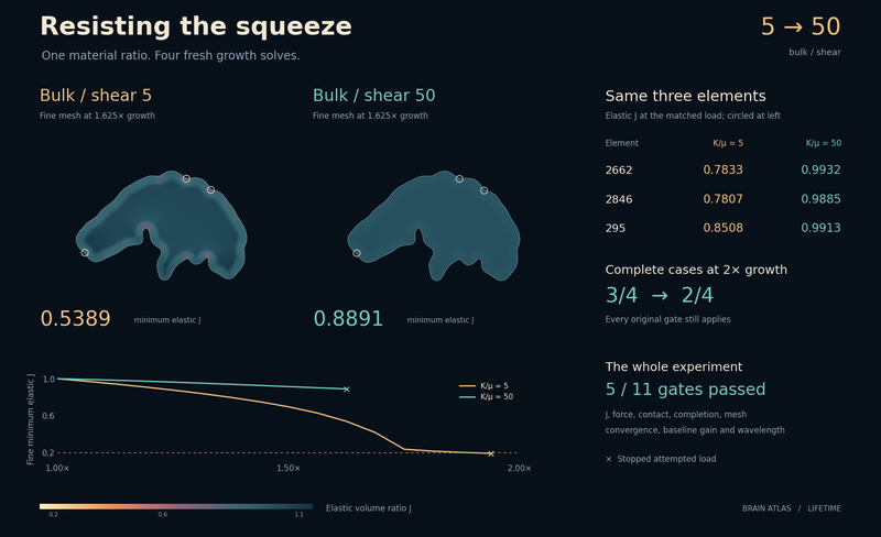
A nonlinear stress test. One material ratio inflates cleanly. The stiffer case stops before the force settles.
MethodsLIFE-010, killed: plane-strain neo-Hookean annulus against frozen radial equilibrium. K/μ=5 completes both meshes; fine L2 error 0.125% versus linear 48.98% and zero 100%. K/μ=50 stops at p/μ=0.05/0.10 on objective change with force Linf 1.97e-7/7.36e-7 above 1e-7. Only 2/4 cases complete; 8/15 gates pass. No fetal geometry, growth or contact; this does not diagnose earlier fetal stops. LIFE-004/006 stay killed. SciPy 1.15.3; annulus-fe note. · derived
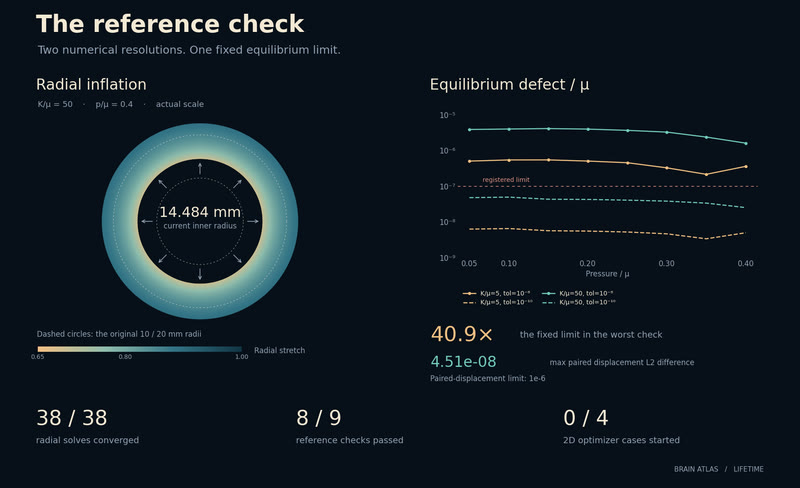
The reference check. All 38 radial solves converged. Two reference checks, at 1e-8 and then 1e-12, stopped the finite-strain experiment before the 2D solver ran.
MethodsLIFE-008 and LIFE-009, killed at the reference gate: plane-strain neo-Hookean radial annulus, K/μ=5/50, p/μ=0–0.4. All 38 BVPs converged, but LIFE-008's 1e-8 checks failed the 1e-7μ defect limit. LIFE-009 tightened the check to 1e-12: defects passed on obtained fields, but the 4,097-node cap stopped two solves and only 14 of 16 pairs existed. No 2D optimizer ran; neither diagnoses fetal stops. SciPy 1.15.3; annulus-finite/precision research notes. · derived
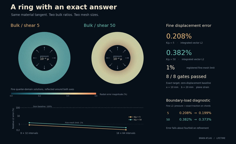
A ring with an exact answer. An exact pressure solution checks the folding model’s tangent: 0.208% and 0.382% fine-mesh error.
MethodsLIFE-007, survives: the unchanged neo-Hookean tangent, solved as plane-strain linear triangles on a synthetic annulus at K/μ=5/50. Fine L2 errors 0.208%/0.382%; all eight gates pass, versus zero at 100%, with error falling ~4× on refinement. Direct LU removes nonlinear stopping here. This small-strain test neither validates fetal folding nor rules out locking elsewhere; LIFE-004/006 remain killed. Bower §4.1.9; annulus-tangent research note. · derived
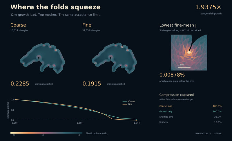
Where the folds squeeze. Three fine-mesh elements cross the compression limit; a trivial layer map matches the localisation score.
MethodsLIFE-005, killed: coarse and growth-only maps both capture 100% of the fine compression deficit in 10% of area; required advantage fails. Neo-Hookean/IPC plane-strain fields on a 21-week dHCP WM contour. Three fine elements have J<0.2; LIFE-004 remains killed. Known-failure diagnosis at g=1.9375, assigned layers. Tallinen doi:10.1038/nphys3632; atlas doi:10.12751/g-node.qj5hs7; IPC doi:10.1145/3386569.3392425. · derived
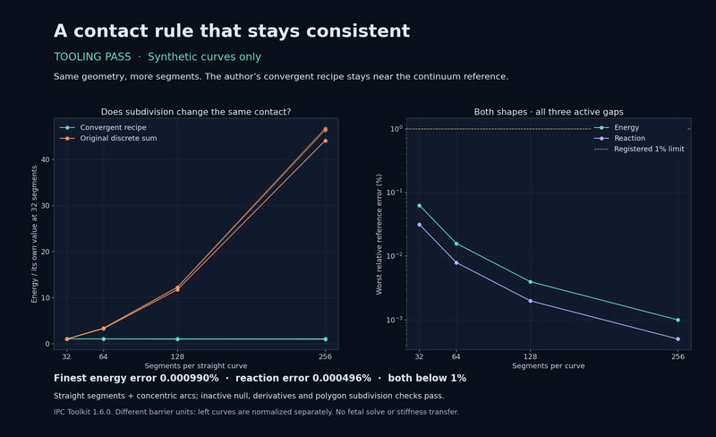
A contact rule that stays consistent. The same curves, split more finely: a weighted contact rule preserves their energy and force.
MethodsTooling pass on synthetic curves: IPC Toolkit 1.6.0's weighted, improved contact recipe matches reference energy and reaction within 0.000990% at 256 segments. The original discrete sum changes by 276–281% on the last refinement. All null, derivative and polygon checks pass. Fixed geometry only: no fetal solve, stiffness transfer or diagnosis of LIFE-020b. Convergent IPC (2023); LIFE-020c tooling note. · derived
A film, through a model of cortex. TRIBE v2 predicts a cortical response to 60 seconds of Big Buck Bunny. The separate TWIN-001 test failed its pre-set check against measured brain responses: the face-map correlation was −0.38, below the required range of 0.59–0.69.
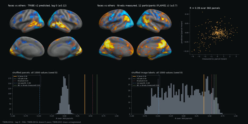
Simulated vs measured face maps: FAIL as registered. We compared TRIBE v2's simulated response to faces versus other images with a measured fMRI map from 12 NI-edu participants. The maps' spatial correlation across 360 cortical regions is R = 0.39. TWIN-001b: FAIL as registered. It missed the required R = 0.54 and did not beat the shuffled-image-label baseline.
MethodsTRIBE v2, average-subject mode, unchanged IBC fLoc image predictions at lag 0; measured NI-edu/OpenNeuro ds003965 v1.0.0 FLAME1 face>others group z-map; fsaverage5 projection and 360 HCP-MMP1 parcels, medial wall excluded. · derived
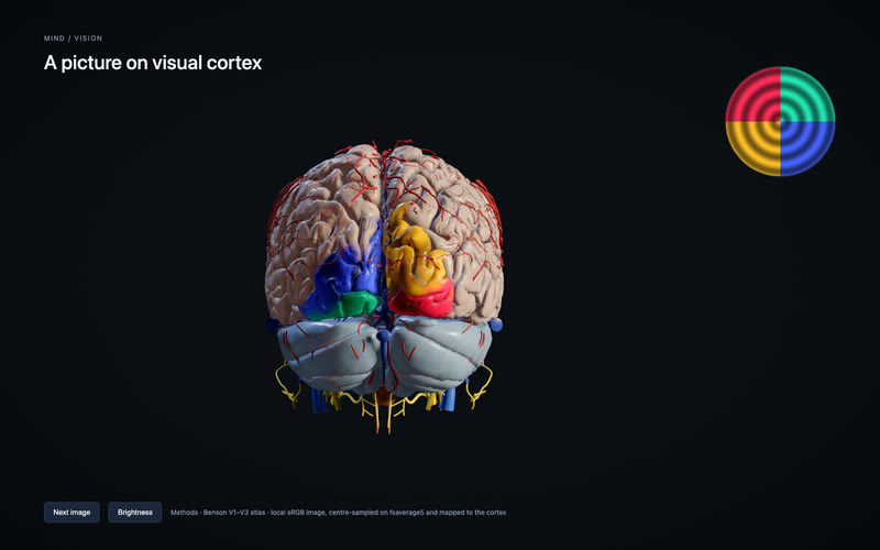
Traveling waves on V1–V3. Rotate a wedge or expand a ring and watch its template projection move across visual cortex.
MethodsTemplate stimulus projection. A synthetic wedge or ring is sampled at Benson 2014 predicted V1–V3 receptive-field centres on fsaverage5. Luminance drives the cortical overlay. The 30° wedge turns once per 12 s; the 1° ring expands from 0.5 to 11.5° in 8 s. One local 256×256 source, steady white with no contrast reversal. Edited screen capture of Play; no neural-response or timing claim. · derived
Paint the visual field. Draw a white stroke and watch its V1–V3 template projection follow.
MethodsTemplate stimulus projection. Locally drawn white strokes in a 24° field are sampled bilinearly at Benson 2014 predicted V1–V3 receptive-field centres on fsaverage5. The existing luminance layer draws the result. Edited screen capture of app 8f87c443 / engine 38410f9b: right-field stroke, left-field stroke, Clear and Back to the bar. Synthetic drawing only; no camera or pixel uploads. · derived
Spatial checks pass; timing not scored. Eight synthetic moving-bar conditions pass. The priority gate leaves performance unscored.
MethodsSpatial checks pass in 8/8 conditions; timing not scored because a Chrome renderer remained at PRI 4. Template stimulus projection through the real Mind camera path: Benson V1–V3, 710 centres, independent atlas oracle. MAE 0.00143–0.00166 versus 0.00784; >99% below zero and best constant. Fake device only. This 8 s clip is a synthetic presentation replay, not measured live throughput. No neural-response or gaze claim. · derived
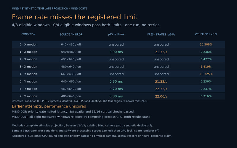
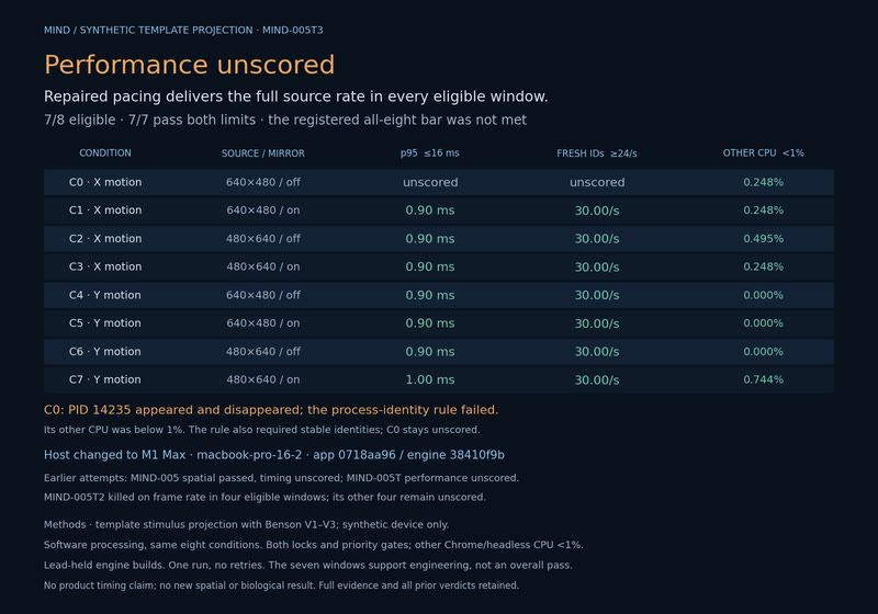
Frame rate misses the registered limit. MIND-005T2: 4/8 eligible, 0/4 eligible pass. Earlier MIND-005 and MIND-005T performance remains unscored.
MethodsTiming hypothesis killed: four eligible windows deliver 21.33–22.33 fresh frames/s (<24); p95 0.70–0.90 ms passes 16 ms. Benson V1–V3 template stimulus projection, fake device only. Four other windows unscored. Earlier performance unscored: MIND-005 (PRI 4; spatial passed), MIND-005T (CPU interference). E2e then GPU locks; unchanged <1% CPU and priority gates. One T2 run, no retry or spatial rescore. · derived
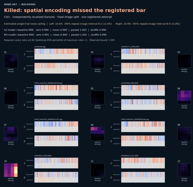
Killed: spatial encoding misses the bar. MIND-007: every model-to-baseline error ratio sat at 0.99–1.00 against a required ≤0.90, on one participant and a fixed twelve-image panel.
MethodsMeasured BOLD5000 Release 2 fMRI from CSI1; independently localized EarlyVis, all 495 voxels. Fixed Gabor-energy ridge; 3301 fit/500 validation/1000 test. Zero, mean, pooled-energy and shuffled baselines; all eight MSE ratios must be ≤0.90 and bootstrap max-ratio bound <1. Repeat-only ceilings LH 10.6%, RH 10.9%. Twelve fixed test IDs; one tagged run. Upstream GLMsingle is not train-only. · derived
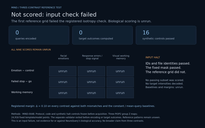
Not scored: input check failed. PIOP2 remains unseen for outcome scoring; the registered input gate stopped the comparison.
MethodsInput gate failed on reference-grid isotropy; biological scoring is unrun. Separate validation stopped before target intensities, query encoding or outcomes. All nine scores, baselines and margins remain unrun. Three fixed PIOP2 contrasts, 24,916 anatomical/model points, Δ=0.10; 16 synthetic controls passed before acquisition. The reference remains unseen for outcome scoring. MIND-004B. · derived
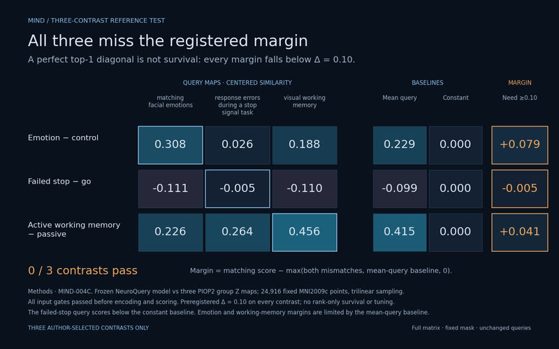
All three miss the registered margin. A perfect top-1 diagonal is not survival: all three miss Δ=0.10.
MethodsFailed the preregistered Δ=0.10 margin on all three contrasts. A perfect top-1 diagonal is not survival. NeuroQuery vs PIOP2 group Z maps at 24,916 fixed MNI2009c points, trilinear sampling. Margins above mismatches, query mean and zero: +0.079, −0.005, +0.041. Three contrasts only; independence conditional on documented publication history. MIND-004C. · derived
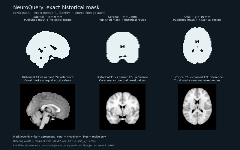
The word map’s reference space. The historical NeuroQuery mask is exact, and its T1 template matches named MNI152NLin6Asym at every voxel.
MethodsSource-lineage audit passes: the historical NeuroQuery/Nilearn recipe reproduces all 28,542 mask voxels; all 902,629 T1 voxels match named MNI152NLin6Asym after lossless reorientation. Zero/one/shift controls differ. The separate binary mask differs at 30 source voxels and is not substituted. Literature-peak registration, cortical projection and biological accuracy remain untested. MIND-003A. · derived
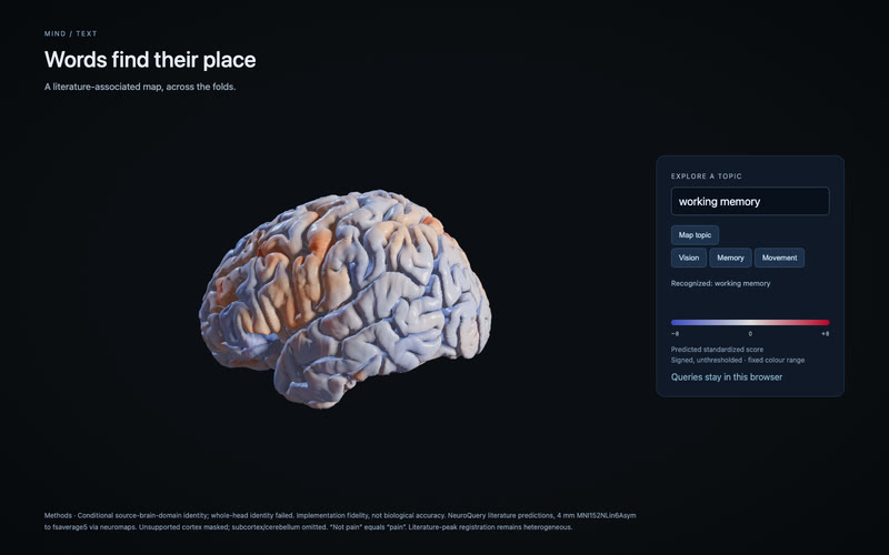
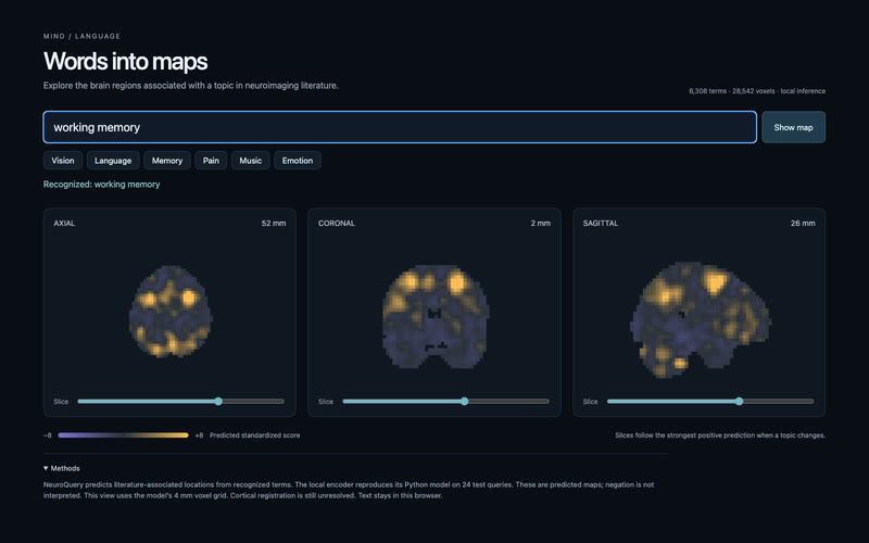
Words into maps. A topic becomes a predicted literature map, computed locally from 6,308 terms.
MethodsImplementation fidelity, not biological accuracy: all 24 queries match the pinned Python encoder (8.32e-8 RMSE). NeuroQuery runs locally in 25.68 MiB. Native 4 mm three-slice view; cortical registration remains unresolved. Unknown text clears the map. Negation is not interpreted. · derived
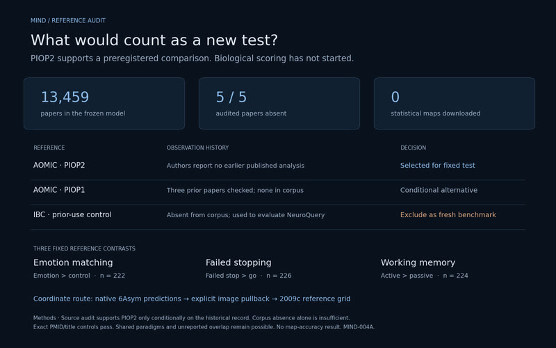
What would count as a new test? A source audit separates a fresh reference candidate from a benchmark already used by the model authors.
MethodsSource audit supports PIOP2 for a preregistered reference test; biological scoring is unrun. Five checked papers are absent from NeuroQuery’s 13,459-row corpus, yet IBC was already used for evaluation. PIOP2 has no earlier published analysis in the author record; unreported overlap remains possible. Three 2009c group contrasts need an explicit template pullback. Failed stop is not successful inhibition. MIND-004A. · derived
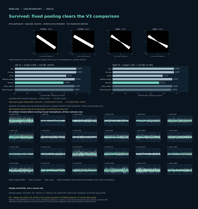
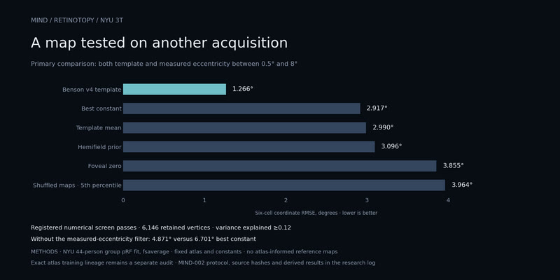
A map tested beyond HCP. NYU 3T: 1.27° error versus 2.92° for the best constant within the matched 0.5–8° field.
MethodsBeats every registered baseline: 1.27° vs 2.92° best constant, with template and measured eccentricity both 0.5–8°. Without the measured-eccentricity filter: 4.87° vs 6.70°. Fixed Benson v4_0 atlas; NYU 44-person 3T group pRF fits (ds003787 1.0.1), six equally weighted V1–V3 cells, variance explained ≥0.12. Exact atlas training lineage remains unresolved. · derived
Localisation one case at a time: a patient's stroke lesion read against arterial territories, Julich-Brain areas and the major tracts.
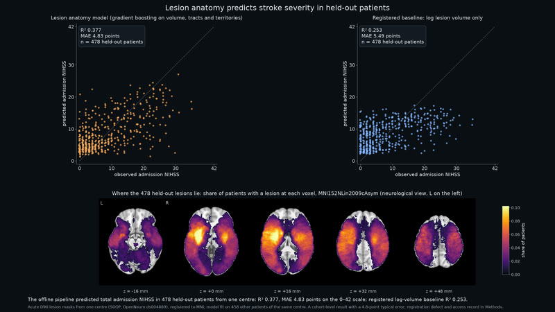
Lesion anatomy predicts stroke severity in held-out patients. The offline pipeline predicted total admission NIHSS in 478 held-out patients from one centre: R² 0.377, MAE 4.83 points on the 0–42 scale; registered log-volume baseline R² 0.253.
MethodsLEARN-001, registered test on SOOP (OpenNeuro ds004889): gradient boosting on log lesion volume, 64 HCP1065 tract loads and 30 Liu 2023 territory loads, fit on 458 development patients (odd ids) and applied once to 478 held-out patients (even ids; 486 masks, 8 excluded by a gross-failure-only registration QC). Acute DWI lesions registered to MNI152NLin2009cAsym; an antspyx transform-list defect displaced every warped mask by about a voxel (Euclidean centroid distances up to 1.17 mm on six development subjects with the same registration, 1.47 mm on ten re-registered; sample measurements, not bounds), and the result stands on those masks. Development scripts parsed the whole participants table before filtering, so reserved NIHSS strings were in memory before scoring; none entered any fit. One scoring run is supported by the retained record, not independently certified. Browser and painted-lesion performance are development checks only, with no held-out estimate. Predictions here are recomputed from the exported model and match the scored R² and MAE to 1e-6. Cross-family review, four rounds, approved. · derived
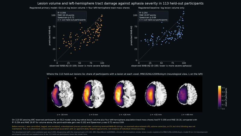
Lesion volume and left-hemisphere tract damage against aphasia severity in 113 held-out participants. On 113 QC-passing ARC reserved participants, an OLS model using log native lesion volume plus four left-hemisphere population-tract mass shares had R² 0.356 and MAE 18.16, compared with R² 0.254 and MAE 19.97 for volume alone; the point-estimate gain was 0.102 and Spearman ρ was 0.72 versus 0.69.
MethodsThe registered point-estimate rule cleared gain ≥ 0.05 and the tract-permutation-null 95th percentile (primary R² 0.356; null p95 0.284), while the 1,000-resample gain interval was 0.037–0.173 and crossed below the 0.05 bar. Before revision 1 was committed, tagged, and receipted, a development process parsed rows containing reserved WAB-AQ strings; no reserved value entered a fit, volume correction, or CV, but strict blinding was not maintained. This is a cohort-level, access-compromised association with an approximately 18-point typical error, not evidence of individual clinical accuracy. The secondary ridge result (R² 0.367) was reported and not judged. · derived
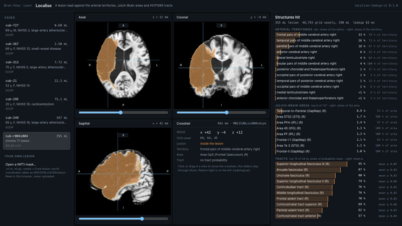
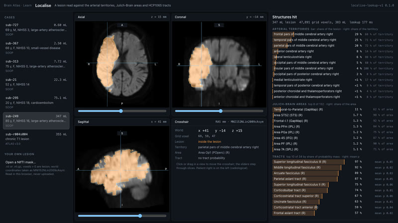
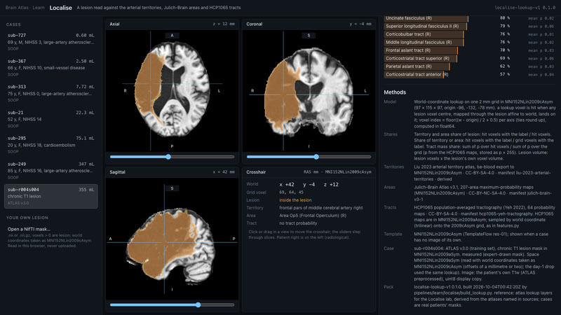
Localise, day 2: the dev page reads a real lesion live. Three slices of the patient's own MRI, the lesion in orange, and the structures it hits: territories, Julich areas and tracts, in under 200 ms in the browser.
MethodsLocalise dev page: a lesion read against the Liu 2023 arterial territories, Julich-Brain v3.1 areas and HCP1065 tracts on one 2 mm MNI152NLin2009cAsym grid, drawn on the patient's own MRI in three radiological slices. Territory and area shares count hit grid voxels; tract loads are the share of each tract's probability mass. Cases: ATLAS v3.0 sub-r004s004 (chronic T1 mask), SOOP sub-249 (acute DWI mask, ANTs-registered). Pack localise-lookup-v1. · derived
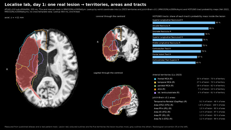
One real lesion, read against the atlases. A 355 mL right MCA infarct from ATLAS lands in three MCA territories and takes most of the right SLF III, arcuate and uncinate.
MethodsATLAS v3.0 sub-r004s004, manual lesion mask and T1w in MNI152NLin2009aSym. World-coordinate lookup into Liu 2023 arterial territories and Julich-Brain v3.1 (MNI152NLin2009cAsym) and the 64 HCP1065 tract probability maps (Yeh 2022, MNI152NLin2009aAsym); no cross-template warp. Tract load is the share of a tract's probability mass inside the lesion. Lookup under 0.5 s on one thread. The clip is the axial sweep. · derived
Glioblastoma treatment margins tested against public recurrence maps: fitted growth models and fixed rules compared at the same treated volume.
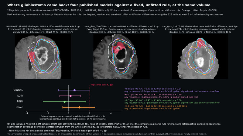
Four published glioblastoma models vs a fixed rule. On 239 included PREDICT-GBM patients (TUM 138, LUMIERE 61, RHUH 40), none of GliODIL, LOTI, PINN or U-Net met the complete registered rule for improving retrospective enhancing-recurrence segmentation coverage over fixed, unfitted diffusion from the whole abnormality. These results do not establish no difference, equivalence, or a true mean gain below +2 pp.
MethodsThis evaluates shipped or reconstructed targets, on this pooled benchmark, at this volume. It does not measure delivered dose, tumour control, survival, other volumes, or newly refitted models. Every target has the voxel count of the reconstructed 15 mm standard target. Fixed, unfitted rule: 2 mm linear diffusion, white:grey 10:1. Registered decision rule, one scoring run, reproduced exactly by a second model family. · derived
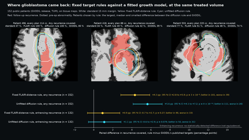
Fixed rules vs a fitted glioblastoma model. At the same treated volume on 152 public glioblastoma patients, a fixed FLAIR-distance rule and an unfitted diffusion rule covered 4.3 and 5.6 percentage points more of the follow-up recurrence than GliODIL's published targets (6.1 with corrected diffusion). On enhancing recurrence there was no statistically detected difference.
MethodsPublic GliODIL release, retrospective (n = 152; enhancing n = 132). Fixed, unfitted rules: FLAIR distance; 2 mm linear diffusion, white:grey 10:1. The diffusion rule averages neighbouring cells arithmetically, allowing spread into cerebrospinal fluid; a registered re-score with harmonic averaging, which blocks flux into zero-diffusivity cells, gave 6.1 points (CI 4.5 to 7.7). Paired Wilcoxon, bootstrap CIs. Before registration, two dry runs scored the plans against the pre-op tumour instead of recurrence; no follow-up label was read. · derived
Corridors to a target, measured against the tracts.
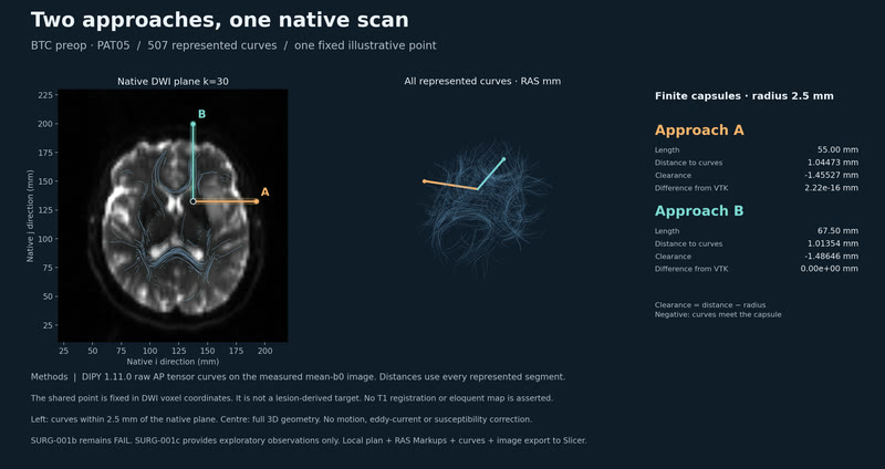
Candidate surgical approaches on a patient's own diffusion scan, scored by their clearance from the reconstructed fibre tracts.
Two approaches, one native scan. Two fixed illustrative paths through 507 reconstructed curves: clearances −1.45527 and −1.48646 mm. Both meet the represented curves.
MethodsSURG-001b remains FAIL. SURG-001c provides exploratory observations only. BTC preop PAT05, DIPY 1.11.0 raw AP tensors; no motion/eddy/susceptibility correction. Fixed illustrative DWI point, not lesion-derived. No T1 registration or eloquent map. Clearance = distance to curves − 2.5 mm radius; always-intersecting ties. Slicer IO: all 552,960 values equal, max physical error 3.63e-6 mm. No tract-truth or outcome test. · derived
A web app on the wgpu engine, built for WebGPU: Explore, Blood flow, Stroke, Seizure, Neurons, Drugs, and now Mind and Stimulate, each with a Methods panel naming its model and data. Free to use.
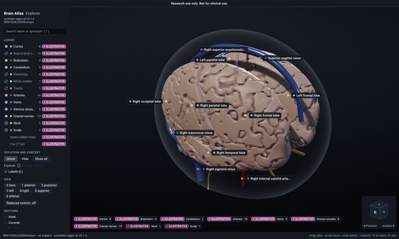
Design screens for all six scenes, built to the component sheet. Layout illustrations drawn from the runs' numbers, not results.
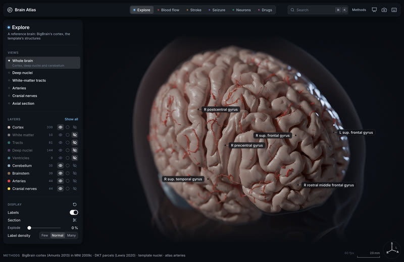
Explore. One real person's brain from their own scans.
MethodsDesign’s screen for the scene, built to the component sheet (tokens 1.3.0, spec 1.1.3): a layout illustration whose numbers are placed from Explore at rest, not a result · Cycles plate behind the chrome, 1470 × 956 at 2× · illustrated
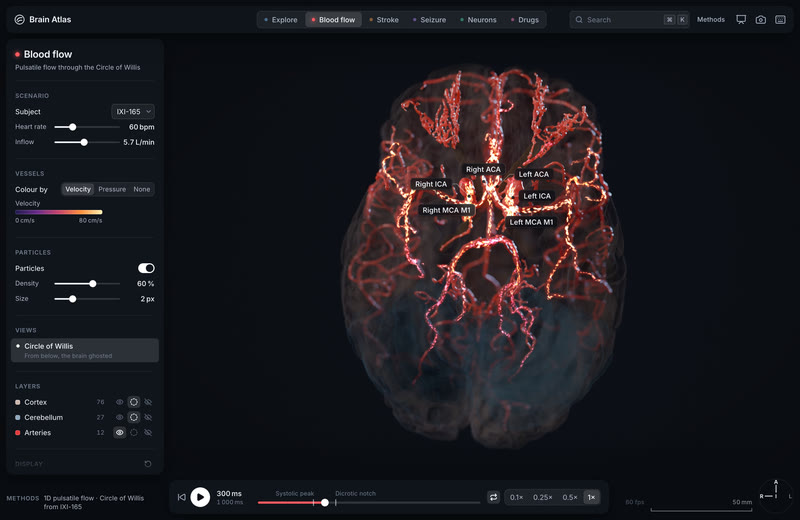
Blood flow. Vessels by velocity, particles, the per-artery readout.
MethodsDesign’s screen for the scene, built to the component sheet (tokens 1.3.0, spec 1.1.3): a layout illustration whose numbers are placed from hemo/ixi165-1d-baseline at 300 ms, not a result · Cycles plate behind the chrome, 1470 × 956 at 2× · illustrated
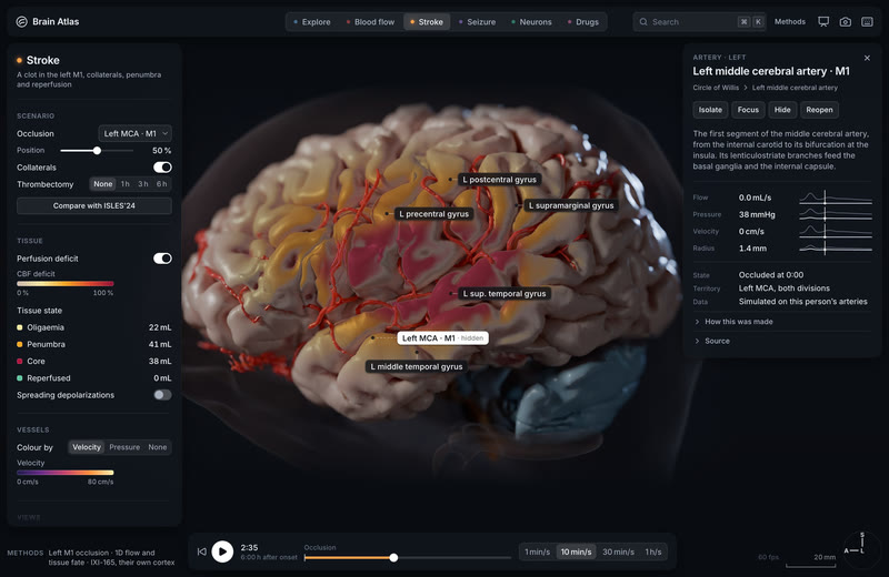
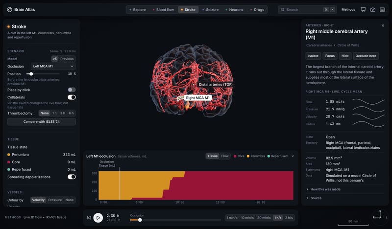
Stroke, 2 h 35 after the clot. Core and penumbra on the clock.
MethodsDesign’s screen for the scene, built to the component sheet (tokens 1.3.0, spec 1.1.3): a layout illustration whose numbers are placed from an illustrative left M1 clot at 2 h 35, not a result · Cycles plate behind the chrome, 1470 × 956 at 2× · illustrated
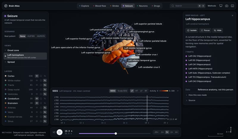
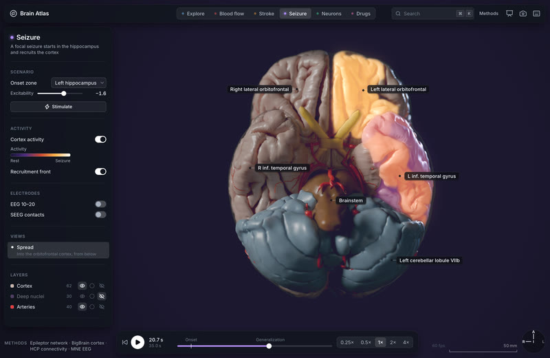
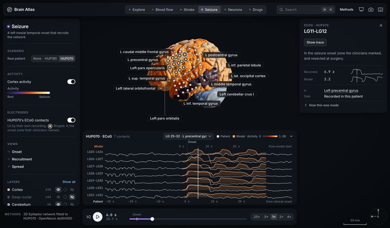
Seizure, spread. The run's last recruitment at 20.7 s, from below: the left orbitofrontal and inferior temporal cortex lit, nothing extrapolated.
MethodsDesign’s screen for the scene, built to the component sheet (tokens 1.3.0, spec 1.1.3): a layout illustration whose numbers are placed from dynamics/seizure-left-mesial-temporal-lateral-spread-v1 at 20.7 s, not a result · Cycles plate behind the chrome, 1470 × 956 at 2× · illustrated
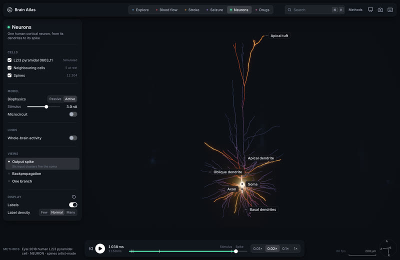
MethodsDesign’s screen for the scene, built to the component sheet (tokens 1.3.0, spec 1.1.3): a layout illustration whose numbers are placed from cells/eyal2018-0603-11-bap-nmda at 1 038 ms, not a result · Cycles plate behind the chrome, 1470 × 956 at 2× · illustrated
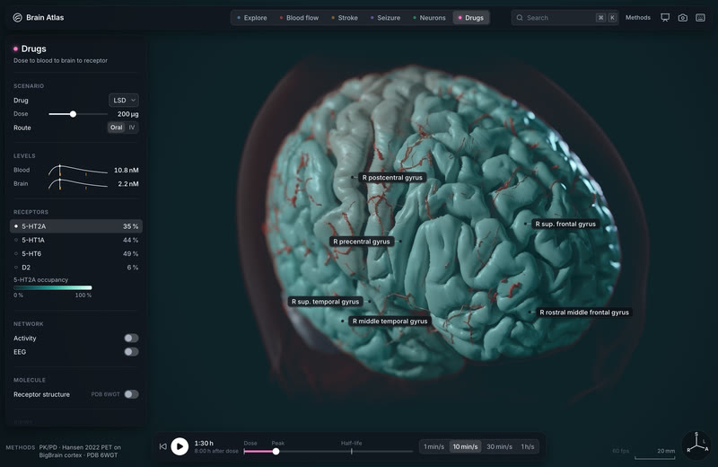
MethodsDesign’s screen for the scene, built to the component sheet (tokens 1.3.0, spec 1.1.3): a layout illustration whose numbers are placed from pharma/lsd-200ug-pk at 1:30 h, not a result · Cycles plate behind the chrome, 1470 × 956 at 2× · illustrated
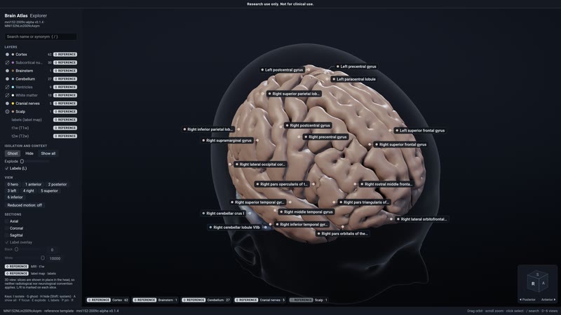
Explore in the native engine. The template's cortex, deep nuclei and cerebellum in the default load state: reference anatomy, an average of 152 adults.
Methodsmni152-2009c-alpha pack: FastSurfer segmentation of the MNI152 2009c template · wgpu engine, native capture, hero camera · reference
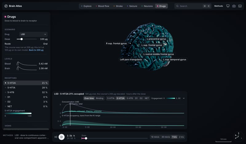
The dose is a slider: LSD's course re-fit live at 100 µg. Slide the dose from the course's 200 µg to 100 µg and the whole course re-fits on its own model: 5.42 nM in blood and 1.08 nM in brain at 1 h 36, 5-HT2A 21 % occupied, 5-HT1A 28 %, 5-HT6 32 %, D2 3 %.
MethodsDrugs: pharma/lsd-200ug-cortex-v1 (oral one-compartment apparent PK, dose to continuous cortex), rescaled live to 100 µg oral on its own model and read at 1 h 36; receptor occupancy from each receptor's Ki range (band on the 5-HT2A trace); drawn by the wgpu engine 42fa1136 (Chrome, 2x) · simulated
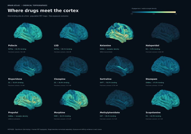
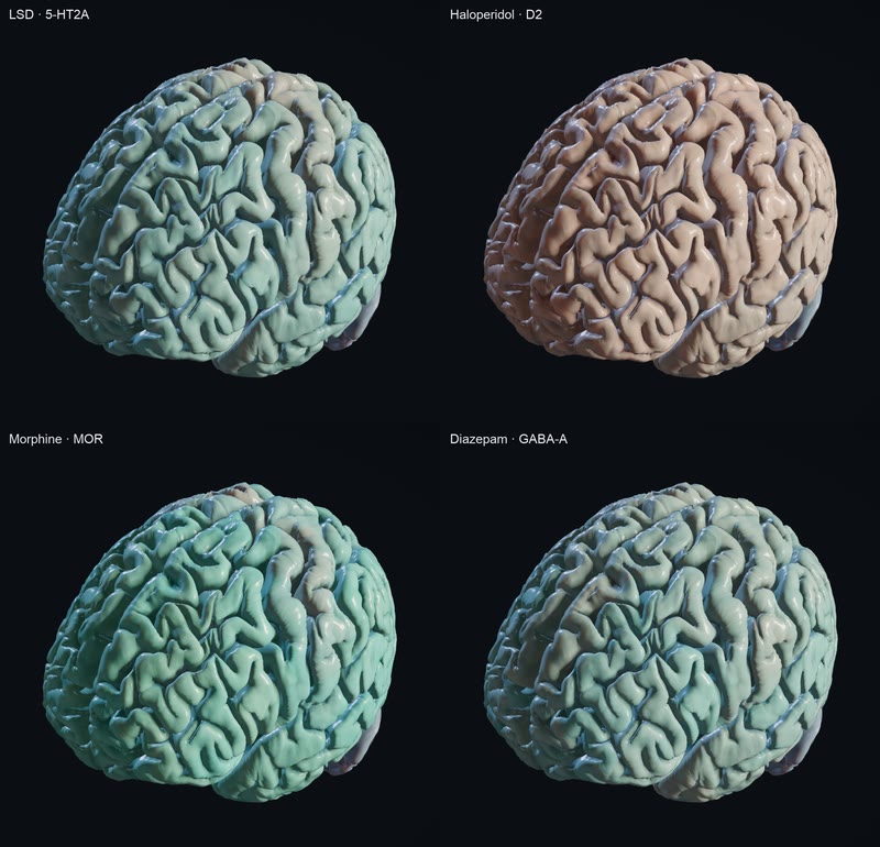
Four drugs on one scale, each where it binds. The app's WebGPU viewport tints BigBrain's cortex vertex by vertex with each drug's engagement, its occupancy times its receptor's PET density there, on one fixed 0–1 scale. Now, under the top of the tint's rise, the colour is the scale's own at 0.30 while the strength still follows the engagement, so weak engagement reads in the drug's hue rather than the tissue's. LSD's 5-HT2A, morphine's MOR and diazepam's GABA-A each carry their colour across the cortex; haloperidol's D2, 10 % occupied, stays a faint wash.
MethodsPharmacology runs on fsLR: LSD's oral one-compartment course (pharma/lsd-200ug-cortex-v1: Dolder 2017 plasma PK, frozen exact-Ki binding, 1:36 h after 200 µg) times Beliveau 2017's 5-HT2A PET surface map; equilibrium site binding for haloperidol, morphine and diazepam (pharma/<drug>) times Hansen 2022's PET receptor atlas. Drawn by engine 2556075c's pharma-sim-v0 reader on bigbrain-cortex-hd through its fsLR map, as design's engagement tint on the fixed 0–1 scale: strength rising from 0.15 to 0.30, a wash up to 0.6 of it under 0.15, and the colour floored at the scale's 0.30 (tokens 0.1.7) (Chrome, 2x) · simulated
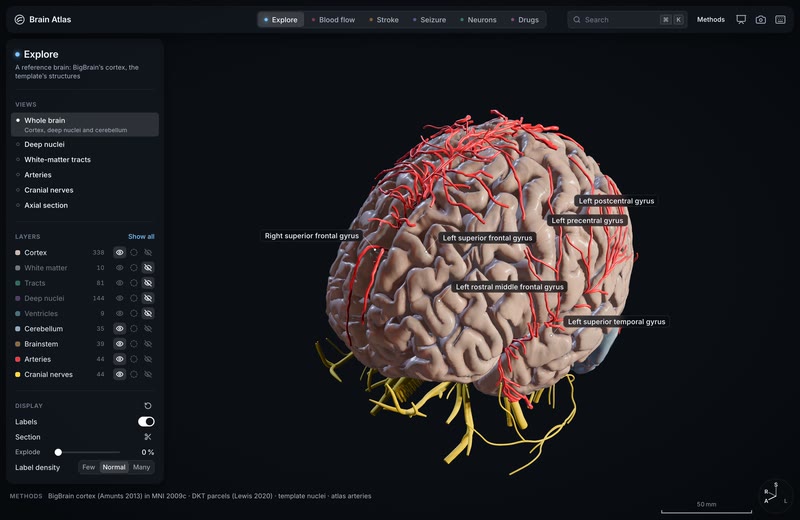
Explore opens on BigBrain's cortex. The app's first scene is now a reference brain: BigBrain's cortex, one person's brain rebuilt from 7,404 histological sections at 20 µm and placed in MNI 2009c, with the template's arteries, cranial nerves, cerebellum and brainstem around it, drawn live in Chrome's WebGPU viewport.
MethodsBigBrain cortex (Amunts et al. 2013), placed in MNI152 2009c by an affine, with DKT parcels per vertex (Lewis et al. 2020); around it mni152-2009c-v0's template structures: FastSurfer's nuclei, cerebellum and brainstem, the Mouches and Forkert arterial atlas (2019) and Z-Anatomy's cranial nerves. Drawn by the wgpu engine 9f964b6 (Chrome, 2x) · reference
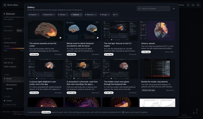
The whole showcase, inside the app. Press G and every card of this gallery appears beside the scene it belongs to: 39 open their exact state in the viewport, the films and figures play with their Methods lines, and each state is a link that carries the scene, the moment and the camera.
MethodsThe app's Gallery sheet: its manifest is generated from the showcase page, its media index and an audit of every card; media play from neurosimulator.ai/media; the links are the app's deep links; the viewport behind is the wgpu engine c45e81fb (Chrome, 2x) · derived
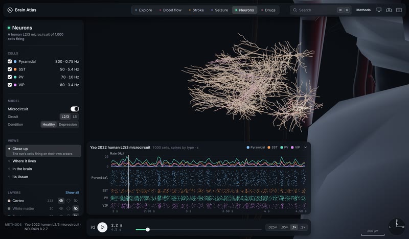
A human L2/3 microcircuit fires on its own arbors. Neurons' Close up in the app's WebGPU viewport: 20 cells of Yao 2022's 1,000-cell human L2/3 circuit, each on its own reconstructed arbor and placed at the H01 site, every compartment's membrane voltage on the activity ramp from −60 to −20 mV. At 2.2356 s two of the drawn cells spike, white-hot at the soma, while the raster below plays the whole circuit's spikes by type.
MethodsYao et al. 2022 human L2/3 cortical microcircuit in NEURON 8.2.7 (cells/yao2022-l23net-baseline, 1,000 cells, 2.0–4.5 s), drawn through its close-up cells/yao2022-l23net-baseline-closeup-r2 (20 cells, 19,457 vertices, 6,616 compartments, dt 0.1 ms) with an illustrative placement that aligns the model's pia with H01's column, by the wgpu engine f1b76b4c (Chrome, 2x) · simulated
H01's real wiring, firing: a cubic millimetre of human cortex in the app. Neurons' Its tissue plays H01's own default run on its 10 s clock: 16,087 neurons of one person's temporal cortex, wired by the 6,526 connections segmented from the electron microscopy, spiking warm through the column's six layers under a resting background drive. The 15,219 driven neurons aren't ringed or flashed cool, so what lights is the tissue itself.
MethodsH01 (Shapson-Coe, Januszewski, Berger et al., Science 2024; Lichtman Lab and Google Research): h01-patch-v0 0.3.0, run h01-lif-resting-recurrent-seed731-v2: exponential-current LIF with phenomenological parameters on H01's eligible neuronal graph, 6,526 of 9,138 connections kept (9,113 synapses; the 104 self-edges excluded), independent Poisson afferent volleys at ALN Temporal_Mid_L E/I rates into 15,219 of 16,087 modelled neurons, seed 731, 949,579 spikes over 10 s; at 4.0 s, drawn by the wgpu engine 42fa1136 (Chrome, 2x) · simulated
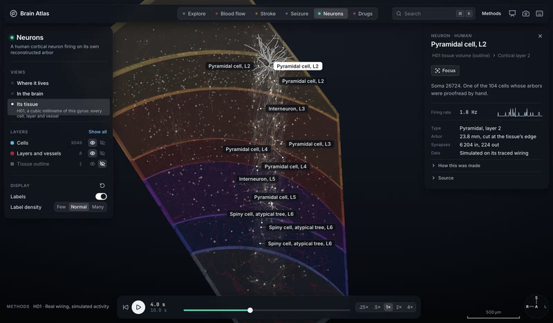
One neuron of H01, read from its own tables: the app's cell inspector. Select any cell in Neurons' Its tissue and the inspector says what it is and how it fired in the run: the column's centre pyramidal cell in layer 2 fires at 1.8 Hz (18 spikes in the run's 10 s), over its rate in each 100 ms, with 23.8 mm of traced arbor that runs out of the imaged tissue and 6,204 synapses in and 224 out. Glia have no rate, and a neuron the run never drives reads Silent.
MethodsH01 (Shapson-Coe, Januszewski, Berger et al., Science 2024; Lichtman Lab and Google Research): h01-patch-v0 0.3.0's neuron table (cell types, 2024 layers, skeleton cable, cut flags, synapse rows) and its default run h01-lif-resting-recurrent-seed731-v2 (exponential-current LIF on H01's eligible neuronal graph, Poisson afferent volleys, seed 731), read by the app from the run's rates.u8, which match its spike counts exactly; at 4.0 s, drawn by the wgpu engine 42fa1136 (Chrome, 2x) · simulated
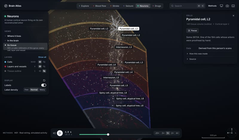
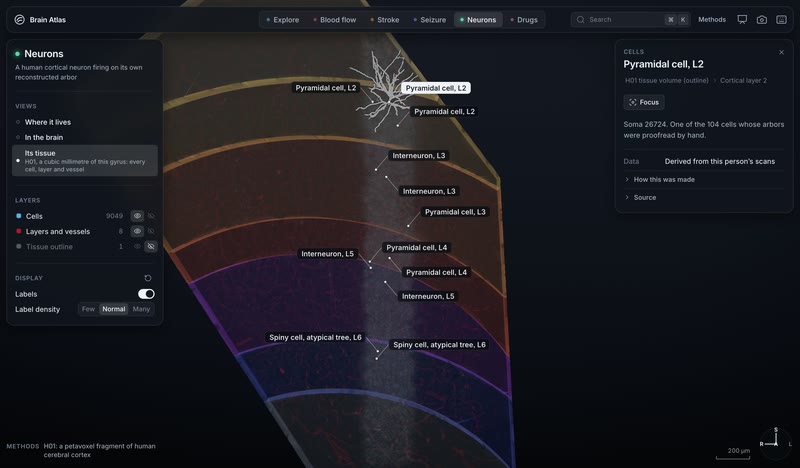
Down to the tissue: H01's cubic millimetre in the app. Neurons' new view opens H01, a cubic millimetre of one person's temporal cortex reconstructed by electron microscopy, in the app's WebGPU viewport: 9,049 named cells (the 104 whose arbors were proofread by hand and the column's 8,945 others), six cortical layers, the white matter and the vessels, with layer-2 pyramidal cell 26724 picked out.
MethodsH01 (Shapson-Coe, Januszewski, Berger et al., Science 2024; Lichtman Lab and Google Research): 4×4×33 nm serial-section EM, c3 agglomeration, 104 cells proofread. Drawn from h01-patch-v0 0.2.1 (neuron table, proofread skeletons, layer and vessel meshes) by the wgpu engine 9f964b6 (Chrome, 2x) · derived
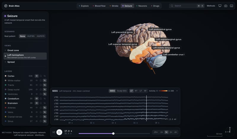
A seizure lights BigBrain's own cortex, live in the app. 17 s into a simulated left mesial temporal seizure, 9 of the model's 82 regions are above a third of its activity range, and the app's WebGPU viewport lights them on BigBrain's real folds (163,842 vertices per hemisphere): the superior temporal gyrus at 98 %, the insula at 74 % and the middle temporal gyrus at 69 %, with the depth electrodes' discharges in the strip below.
MethodsDelayed six-state Epileptor network on HCP-derived connectivity (dynamics/seizure-left-mesial-temporal-lateral-spread-distributed-v1: fsaverage, 82 regions, 1,051 frames over 35 s, OpenMEEG depth potentials), moved onto bigbrain-cortex-hd 0.1.0 through its map-fsaverage by the wgpu engine 9f964b6 (Chrome, 2x) · simulated
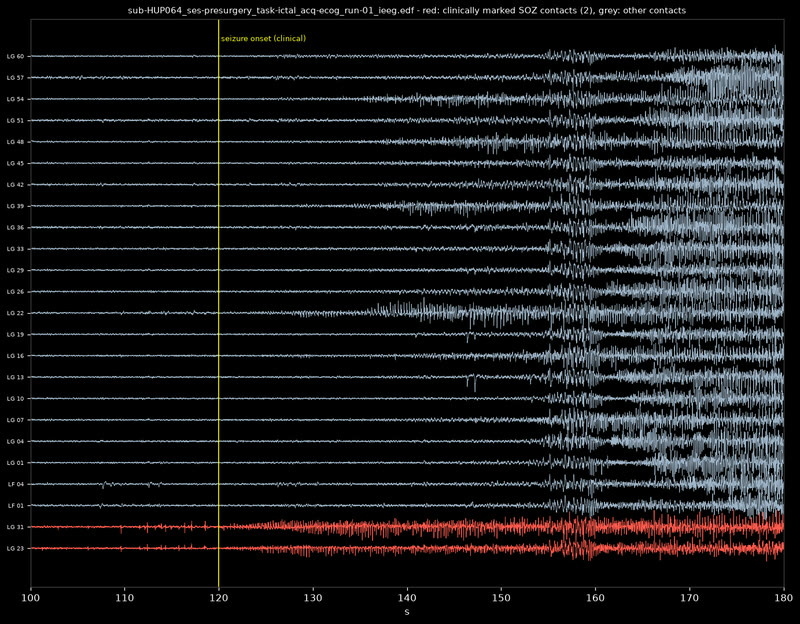
A real patient's electrode, read from their own recording: the app's electrode inspector. Click any electrode site in Seizure and the inspector reads its channel. On HUP070's ECoG grid, LG11-LG12 sits in the left precentral gyrus, in the onset zone their clinicians marked and later resected; at 6 s their recording there reads 6.9 z beside the fitted model's 2.2. On the model run, scalp T7 picks up 212 µV, 48 % of its variance from the left insula: an exact split through the head model's gain.
MethodsHUP070's ictal ECoG (OpenNeuro ds004100 v1.1.3, CC0): bipolar line length as a robust z, the contact pairs' midpoints and the clinicians' onset-zone and resection marks from observation.json, beside the 2D Epileptor network fitted to them (dynamics/hup-seizure/sub-HUP070). The model run's scalp EEG is MNE's fsaverage three-compartment BEM (dynamics/seizure-left-mesial-temporal-lateral-spread-distributed-v1), and each region's share of a channel's variance comes from its gain and source scale. Read by the app; at 6.0 s, drawn by the wgpu engine 42fa1136 (Chrome, 2x) · measured
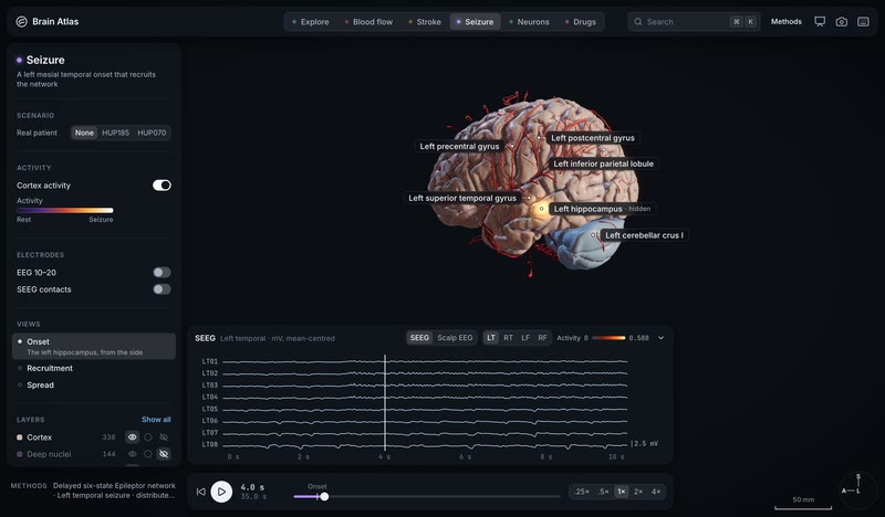
The hidden onset zone glows through the temporal lobe. 4 s into the model's left mesial temporal seizure, the onset zone is the left hippocampus, deep inside the brain. The app's WebGPU viewport lays design's onset glow over the tissue in front of it (15 mm around its centroid, the activity scale's amber at 85 %) on BigBrain's cortex, with BigBrain's own pial arteries draped on its folds.
MethodsDelayed six-state Epileptor network on HCP-derived connectivity (dynamics/seizure-left-mesial-temporal-lateral-spread-distributed-v1), moved onto bigbrain-cortex-hd through its fsaverage map; pial arteries from bigbrain-pial-vessels 0.1.0 (three 7 T TOF networks, Bollmann 2022); the glow is design's onset glow, an illustrative marker of the deep onset; wgpu engine f1b76b4c (Chrome, 2x) · simulated
Any vessel, live: its flow, its territory, and Occlude here. Two and a half hours into a left M1 clot, pick the other M1 and the inspector reads it from the live solve: 1.85 mL/s at 91.9 mmHg and 28.7 cm/s through a 1.43 mm radius, open, feeding the right MCA territory, with Occlude here to put the clot there instead and re-solve the whole network.
MethodsStroke at 2 h 35 after a left M1 occlusion: tissue hemo/ixi165-stroke-v5-l-m1-intermediate-untreated, flow hemo/ixi165-1d-v5-occl-l-m1 with hemo/ixi165-1d-v5-baseline as the reference, spreading depolarizations dynamics/spreading-depolarization-ixi165-m1-v5-841dcfd0-t0-v1; the live rows are hemo-rt's cycle means on the v5 Circle of Willis network (a model circle, not this person's), the territory is each region's share of its blood through this vessel by the reference run's mean flows, mixing fully at each junction; Occlude here moves the clot and re-solves; drawn by the wgpu engine 42fa1136 (Chrome, 2x) · simulated
Every bet writes its kill rule before the data are opened, and the kills stay on the page. So far: geometry and metabolism hold at the map level, the physics checks out, and template networks at n ≈ 50 can't carry mechanistic claims.
Perforator territory, distance from the watershed and large-artery probability predict lesion frequency. The high-extraction mechanism does not (p = 0.916).
MethodsISLES'22 lesion-frequency map (400 cases) × Liu 2023 territories, the Mouches–Forkert artery atlas and neuromaps CBF and CMRO2; BrainSMASH spatial nulls · bet 2 · derived
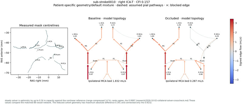
A patient's Circle of Willis physics predicts infarct growth.
ΔR² −0.016 [−0.071, 0.005] for growth over the clinical baseline.
48 strict anterior-circulation ISLES'24 cases in nested cross-validation: the capacity index does no better than a 4-bit variant code. The physics, the 149-case feature table and the site inventory stand.
Methods1D steady solver on each patient's TopCoW circle graph, cross-checked against openBF (rank ρ 0.9997) · bet 1 · derived
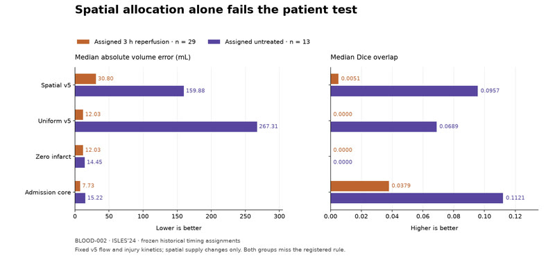
Spatial supply alone predicts a patient's final infarct.
Median volume error 30.8 mL, against 12.0 for zero infarct and 7.7 for the admission core (3 h group, n = 29).
Untreated, n = 13: 159.9 mL against 14.5 and 15.2. IXI-165's v5 supply routed into 42 ISLES'24 cases at fixed injury kinetics, the rule frozen before scoring. v5 itself met its registered rule, and predicting no infarct at all still beat it: 12.7 mL against 37.0.
MethodsPassive route tracing of the v5 flows into each case's own vessel map, 29 assigned 3 h and 13 untreated, scored natively against the ISLES'24 final infarct with uniform, zero and admission-core controls · bet BLOOD-002, research note 2026-10-02-blood-spatial-result · derived
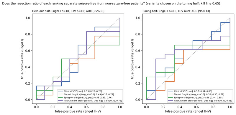
A template Epileptor fitted by simulation-based inference finds the seizure focus.
Held-out AUC 0.55 [0.33, 0.76] against a 0.65 bar.
Neural fragility 0.49, the clinical onset zone 0.53, recruitment order 0.54. Every ranking finds the resected channels (AUC 0.68–0.74); none predicts the outcome in this cohort.
MethodsHUP ds004100, 55 patients split into tune and held-out halves, Engel I vs II–IV · bet 7 · predicted
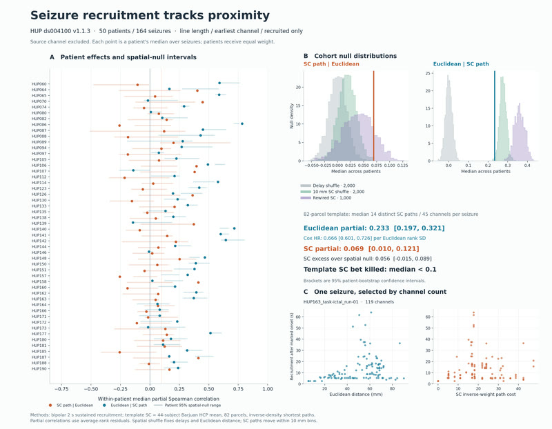
50 patients, 164 seizures: recruitment order follows proximity, with a hazard ratio of 0.666 per distance-rank SD. Patient-specific tractography is untested.
MethodsHUP ds004100 recruitment times × an 82-parcel template connectome; registered plan · bet 9 · derived
The clot's position along M1 decides the deep infarct.
Lenticulostriate bed lost: 30 % proximal vs 13 % distal (p = 0.0026).
In 72 ISLES'24 M1 patients the loss falls with distance from the carotid terminus (ρ = −0.37). Insula and cortex don't differ, so the effect is specific to the end-artery bed.
MethodsISLES'24 training set, clot near-edge measured on CTA · bet ISLES-003 · derived
Killed
A learned surrogate makes 1D cerebral flow real-time.
22.7 % held-out flow error, 13× faster than physics that already solves in 0.85 ms.
The MLP buys no interactivity on this graph: the exact periodic solver already answers in the browser in 1.7 ms. Nonlinear teachers are next.
Methodsa learned MLP surrogate against the exact Rust/Wasm periodic solver hemo-rt · predicted
Won
The engine's cortex matches the reel at its own camera.
Luma quartiles within 0.02 of the Cycles still.
IQR 0.12 to 0.37 against the reel's 0.36; the top 1 % is still short. Killed alongside: the capillary net as colour, which reads as a red honeycomb on the engine.
Methodsengine look bets ENGINE-LOOK-001 and -002 · derived
Measured
Three drugs, three connectivity fingerprints.
Psilocybin +0.107, methylphenidate +0.057, LSD +0.010 global Δz.
35 unique acquisitions from seven people scanned repeatedly, plus 15 LSD participants. The targets for the receptor-informed model are published; the model stage is next.
Methodsds006072 and ds003059, pre-registered plan frozen before the FC computation · bet 3 · measured
Data
Real data at every scale.
55 datasets requested, 46 acquired and verified, each with its own manifest saying where it is and how to fetch it again.
264dataset manifests, each saying where the data is and how to fetch it again
46of 55datasets acquired and verified; the rest wait on access approvals
2.7TBof raw data on the project's store
7,404stained sections in BigBrain, one human brain at 20 µm
503,085streamlines in the HCP1065 tract atlas, 102 M points before simplification
163,842vertices a hemisphere on the canonical cortex, BigBrain's own
57,215typed cell bodies in the H01 pack; its column holds 7,698 skeletons and 21.0 M synapse points
149+ 250stroke cases on disk from ISLES'24 and ISLES'22, plus 1,452 strokes in ATLAS v3.0
57HUP intracranial EEG patients, 208 seizures
19PET receptor targets and 2,579 measured affinities behind the drug scenes
35+ 60rest-fMRI runs behind the drug fingerprints: psilocybin and methylphenidate (ds006072), LSD (ds003059)
1mm³of human cortex in H01 at 4 × 4 × 33 nm, 6.1 GB of release subset on disk
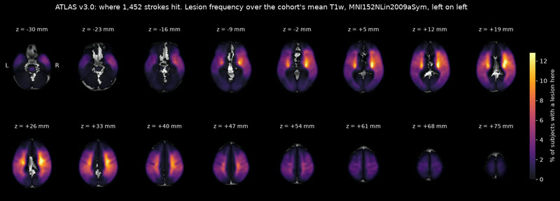
Where 1,452 strokes hit. ATLAS v3.0 lesion frequency; the peak is 15.5 % of subjects, in the right putamen.
MethodsATLAS v3.0 lesion masks, voxel-wise frequency over its 1,452 subjects · measured
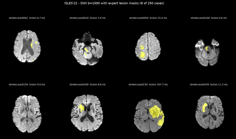
ISLES'22: 250 cases with expert lesion masks. Eight of them on diffusion MRI; the challenge's 400-case aggregate heatmap is what the lesion-geography bet used.
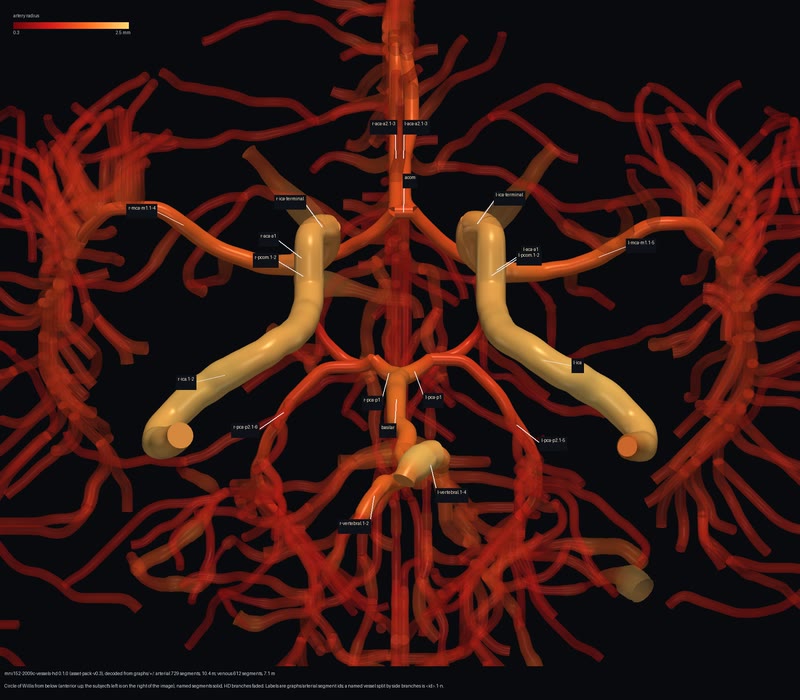
The named circle. vessels-hd: 729 arterial segments (10.4 m of vessel) and 612 venous (7.1 m) on the template, the Circle of Willis labelled, seen from below.
Methodsmni152-2009c-vessels-hd 0.1.1, decoded from the pack's graphs · reference
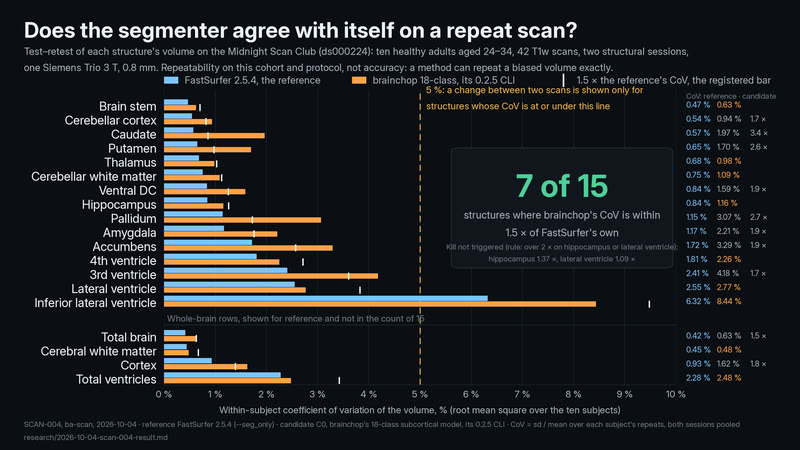
Does the segmenter agree with itself on a repeat scan? On ten adults scanned 42 times, FastSurfer repeats a structure's volume to 0.4–2.6 % and brainchop stays within 1.5 × of that on 7 of 15 structures: each method's agreement with itself, not its accuracy.
MethodsSCAN-004, repeatability not accuracy: within-subject CoV per structure on MSC ds000224 (10 adults, 24–34, 42 T1w scans, 2 sessions, one Trio 3 T, 0.8 mm). Reference FastSurfer 2.5.4 --seg_only repeats to 0.42–2.55 % (inferior lateral ventricle 6.3 %); candidate C0, brainchop's 18-class model run by its 0.2.5 CLI with the page's weights, within 1.5 × of that on 7 of 15; kill not triggered. Self-agreement cannot see a bias against truth. · derived
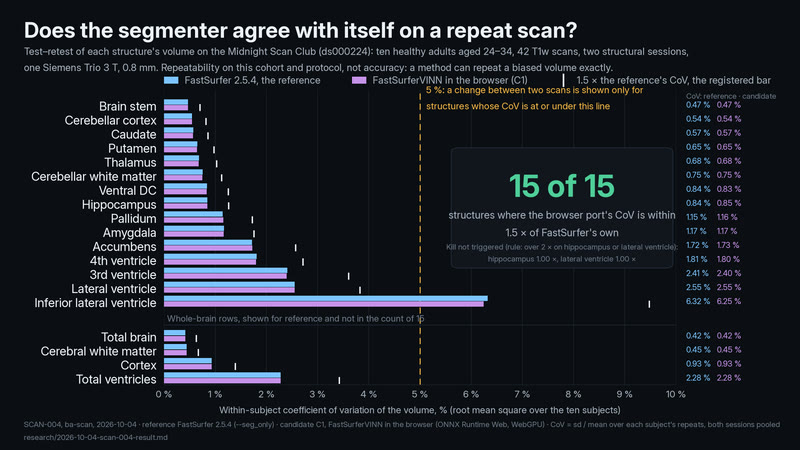
In the browser, FastSurfer repeats as well as it does natively. On 42 scans of ten adults, FastSurferVINN running in the browser is as repeatable as FastSurfer 2.5.4 on every structure (CoV ratio 0.99–1.01; 15 of 15 within the registered 1.5×). It is the same network on another runtime, so this shows the port is faithful, not that it is accurate.
MethodsSCAN-004, repeatability not accuracy: within-subject CoV on MSC ds000224 (10 adults, 42 T1w scans, one 3 T scanner, 0.8 mm). Candidate C1, FastSurferVINN in the browser (ONNX Runtime Web, WebGPU), matches FastSurfer 2.5.4's CoV on every structure (ratio 0.99–1.01, 15 of 15 within 1.5×, kill not triggered); its labels differ from the reference's on every scan (410–641 voxels). Same network, another runtime: port fidelity, not an independent estimate. · derived
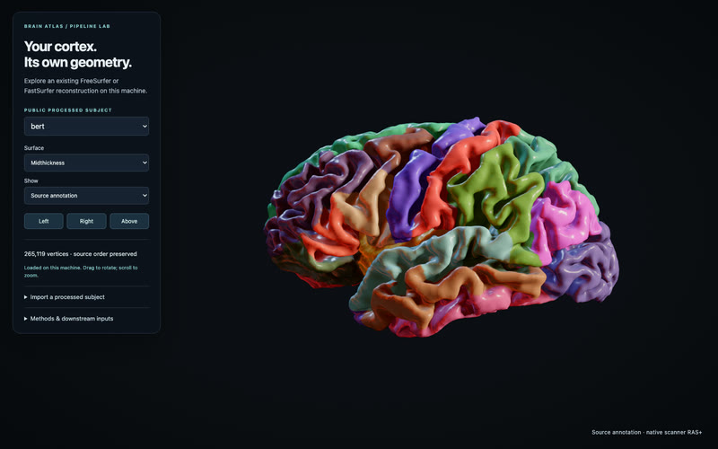
Your cortex, in its own geometry. An existing reconstruction becomes a native 3D brain, preserving its vertices, labels and thickness.
MethodsPIPE-001: FreeSurfer bert processed cortex, imported with the full scanner-from-tkr affine. All 265,119 vertices retain their order; arrays and topology match independent readers exactly. Import coordinate error <0.0001 mm on IXI165, bert and MNE sample. Upstream segmentation accuracy is not measured; Lifetime, Stimulate and Stroke need additional inputs. · derived
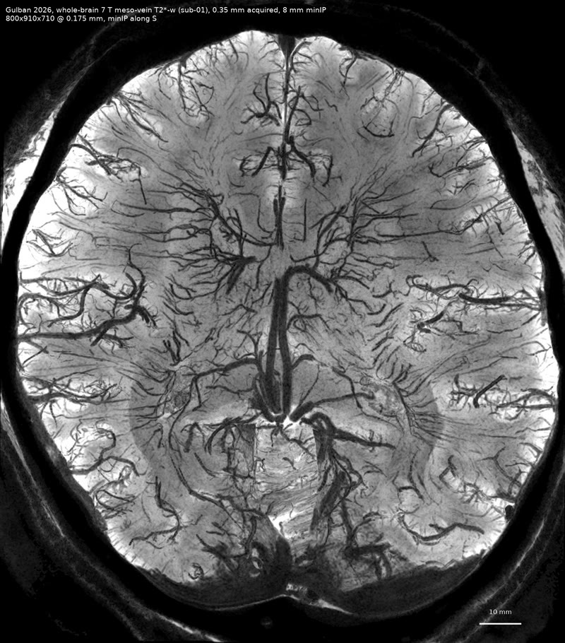
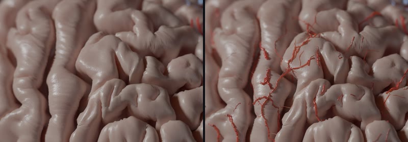
Real pial arteries on BigBrain's cortex. The same crop of the Explore hero without and with the 7 T pial arteries (Bollmann 2022) draped onto BigBrain's folds: 26 m of real vessels in one network, now a published pack.
MethodsThree 7 T time-of-flight acquisitions (Subject_03, Subject_01, twoEchoTOF) fitted to BigBrain's pial surface in MNI 2009c by a trimmed iterative closest point, every centreline point moved to the nearest surface point and lifted by its radius, the three feathered into one network by leaf pruning. A drape, not a registration: the vessels are real, their course on these folds is not. Cycles, living look, 2940×1912, 256 spp. · derived