Brain Atlas / 2026-10-12 / corrected 13 October / Methods
The platform, today · 12 October
69.433 seconds · 1920 × 1080 · 30 frames per second · silent
Corrected 13 October 2026. The original reel's glioma-growth excerpt at 62–71 seconds drew tumour density through a displacement field that did not match its mass model. That excerpt is removed, including its dissolves and an additional frame at each boundary. The faulty images have not been replaced by a new model result.
The remaining ten scenes retain their order, captions and source images. They are separate studies with different clocks, populations and baselines, not one coupled simulation. The film remains silent, and source images are fitted without cropping. Existing dissolves remain; the removal join is a hard cut.
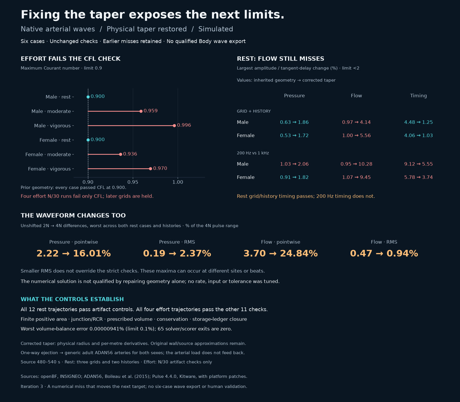
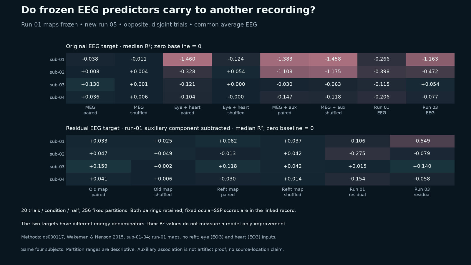
The larger running model has not passed its learning check, all six corrected-taper cases still miss overall, and no disjoint-trial EEG range clears all three baselines. The ultrasound result is one water control. Toy listeners are not people and chromosome partitioning remains incomplete. The ribosome comparison keeps its stricter tomogram-count disclosure.
The remaining glioma recurrence simulation draws no postoperative mass effect and is not fitted to a patient. Its fluid spaces take no tumour cells. Its contextual template anatomy is distinct from the two brain-only measured scan displays.
Source playback seconds and simulated time are different. The listed reel intervals include surviving dissolves, so neighboring scenes can overlap. Displayed interval endpoints are rounded to the nearest millisecond from the 30-frame-per-second edit.
01 / 0–9 s · real data + illustrated
One scan becomes a glass brain
Precision Aging Network · Barnes, Huentelman, Chen et al. / OpenNeuro ds007522 2.0.1 / FreeSurfer 7.4.1
A defaced T1-weighted scan, sub-HML0441 session 01, from the Precision Aging Network (Barnes, Huentelman, Chen et al.; OpenNeuro ds007522 2.0.1). FreeSurfer 7.4.1 recon-all supplies aseg segmentation, pial surfaces and Desikan–Killiany regions. Smoothed marching-cubes structure meshes and glass surfaces are drawn in Blender 5.2 Cycles. The colours, transparency and camera are illustrated; this is a segmentation display of one measured scan, not simulated brain activity. The film shows brain tissue only, with no facial surface. This 12–21 s excerpt preserves the source frame and its annotations.
Source film · Source playback 12–21 seconds.
Source file identity
SHA-256 cdf455ab99bee34f1c6563e632e0c41a411492ecfbc6595c77ed9bfe9b0fa83113,885,396 bytes
02 / 8.5–14 s · real data + simulated + illustrated
More runners; the learning check still misses
Carter et al. 2024 / AddBiomechanics · Werling et al. 2024 / MyoSuite · MuJoCo · MuscleMimic
MyoFullBody in MuJoCo 3.4, fine-tuned from MuscleMimic mm-10m-2. A 12-runner controller and a 33-runner one re-enact held-out runner P017 against measured force plates (Carter et al. 2024 via AddBiomechanics 1.0, Werling et al. 2024). The same training budget gives each new runner roughly a third of the earlier training. The two 33-runner seeds follow the new runners at slopes +0.17 and +0.23, short of +0.66. The run with 11.9 mm held-out bounce error failed its learning check; those held-out numbers are exploratory. One seed beats the constant-bounce baseline of 13.6 mm, the other at 13.8 mm does not. The held-out follow slope +0.16 (SE 0.09) is not above zero by two standard errors. This does not establish whether more runners help. Simulated knee loads remain about 15 body weights. Generic bones, head omitted.
Source film · Source playback 0.25–5.75 seconds.
Source file identity
SHA-256 34cd9a2e91d1e1d4d6fd561fb230561f162956348ed3bb81cf89c437a2182afb1,005,601 bytes
03 / 13.5–20.5 s · simulated
The taper is fixed; six cases still miss
openBF · INSIGNEO / ADAN56 · Boileau et al. 2015 / Pulse 4.4 · Kitware
Corrected physical taper in a one-way arterial model using openBF (INSIGNEO), ADAN56 (Boileau et al. 2015) and Pulse 4.4.0 (Kitware, with platform patches). The T adapter, inputs and strict 2% comparison limits are unchanged; source time 480–540 s. Rest grid/history timing now passes, but flow-amplitude changes reach 4.14% male and 5.56% female. At 200 Hz, flow changes reach 10.28% and timing 5.55%; male pressure misses at 2.06%. All four effort cases exceed CFL 0.9 (up to 0.996), so their later grids are held. Conservation, finite area, junction/RCR, prescribed volume and ledger controls pass on all 16 completed trajectories. All six cases still miss overall, with no qualified export. Generic adult geometry is used for both sexes. The full figure is fitted without cropping.
Source image · The full source image is fitted without cropping.
Source file identity
SHA-256 b4a100eb734f68dffde9c64dc02f210031844fcf85ca0ed4982368907ef4420a275,301 bytes
04 / 20–26 s · real data + simulated
Some same-trial signals carry; disjoint trials do not
OpenNeuro ds000117 · Wakeman & Henson 2015
Aggregate EEG/MEG/eye/heart statistics and statistical predictions, using four reused development subjects. Run-01 maps are frozen and tested on run05 faces minus scrambled, 0–400 ms. Original-EEG MEG sign stability carries in 19/24 same-trial cells and 0/24 disjoint cells; no disjoint range clears all three baselines. Baselines are zero and run01/run03 EEG. The 5/10/20-trial partitions overlap; their descriptive ranges are not confidence intervals or an independent subject test. Original and residual R² use different denominators, and auxiliary-channel subtraction may remove neural signal. The source card uses the public label simulated for its statistical predictions; the recordings are real data.
Source image · The full source image is fitted without cropping.
Source file identity
SHA-256 c08b52dcef8eaced95446dd50f6286237b19ea5a74ebeeefe4cae052e48dfba8165,286 bytes
05 / 25.5–31.5 s · simulated
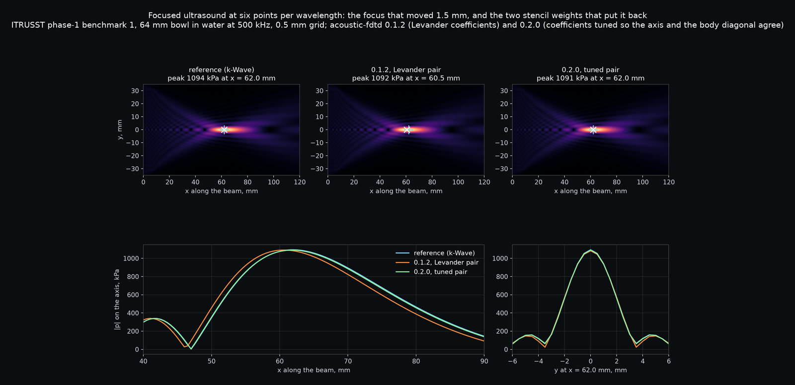
Two weights bring the focus back—in water
ITRUSST phase 1 · Aubry et al. 2022 / k-Wave reference / Brain Atlas FDTD 0.2.0
ITRUSST benchmark 1: a 64 mm bowl in water at 500 kHz. At a 0.5 mm grid (six points per wavelength), FDTD 0.1.2 put the focus 1.5 mm short of the published k-Wave reference and missed four of six predeclared bands. The fourth-order scheme's two stencil weights are now tuned per run so axial and body-diagonal waves agree at the drive frequency. In this control, maximum direction error falls from 0.47% to 0.05%; the peak returns to the reference cell and all bands pass. This is one water control, not a claim about skulls at 0.5 mm. The old-weight GPU-pod control matches the earlier Mac run to every printed digit and repeated runs are byte-identical. Source: ITRUSST phase-1 benchmarks, Aubry et al. (2022), Zenodo 6020543; reference k-Wave at 0.1 mm.
Source image · The full source image is fitted without cropping.
Source file identity
SHA-256 a0ff145bfbe2e8ddfcb08c188fc906045b84a715636d4eb7f9d1351b4c3e3276132,458 bytes
06 / 31–39 s · simulated + illustrated
Toy listeners follow a sound field
Room reflections · Allen & Berkley 1979 / SADIE II KU100 / CARFAC v2 / Brain Atlas 2026
A scripted four-metre room: 1,500 Brownian agents walk a sound-level gradient toward a wanted level. They are toy listeners, not a model of people. The shared field adds direct sound and first-order images in four walls, absorption 0.1, following Allen and Berkley (1979). It sums energy only, without phase, higher-order reflections or a reverberation tail. The source has binaural sound through SADIE II KU100 head-related transfer functions and CARFAC v2, with speech from LibriVox; this reel removes all audio. The producer recaptured the scene with walls on, correcting the earlier dropped blank frames. Drawing and dots are illustrated; no human anatomy is rendered.
Source film · Source playback 16–24 seconds.
Source file identity
SHA-256 e845a1d865cba4b75e256cec58c6c806784142751dc6bbb7778081c1df4c0ffd5,566,048 bytes
07 / 38.5–46.5 s · simulated + illustrated
Two chromosomes begin to part
4D whole-cell model · Thornburg et al., Cell 2026 / btree_chromo / polymer-bd 0.4.1
The chromosome model polymer-bd 0.4.1, a WebGPU port of btree_chromo, starts from the state reached at 3,924 simulated seconds in cell 35. Over 75 four-second windows to 4,224 s, directives come from the 4D whole-cell model’s own writer (Thornburg et al., Cell 2026), driven by its published cell-35 membrane calls and counts. The neck deepens and the two daughter chromosomes collect toward opposite lobes. Beads per lobe and partitioning are read by the model’s own check: partitioning is not complete. Colours by daughter and the camera are illustrated. The video’s playback time is not the model’s clock; this excerpt keeps its source annotations.
Source film · Source playback 10–18 seconds.
Source file identity
SHA-256 8d1ac9c2a0aca7c452fd479c227f74ba85eb55515b4e2b5f1ef5f1c5a209973015,403,146 bytes
08 / 46–55 s · real data + illustrated
Ribosome pairs in their own tomogram
JCVI-syn3A · CZ CryoET Data Portal 10442 / EMD-75898 / PDB 7P6Z
One cryo-electron tomogram of JCVI-syn3A, CZ CryoET Data Portal dataset 10442, mirrored for display to match the ribosome map’s hand. Template matching with pytom-match-pick 0.14.0 and EMD-75898 followed by refinement gives 21 pairs among 193 ribosomes in this tomogram. Pair criteria: 20–35 nm separation and within 30 degrees of the first cell’s pair pose. The displayed atoms are PDB 7P6Z, a Mycoplasma pneumoniae 70S model fitted into EMD-75898, not syn3A density. Camera, fades and colours are illustrated. The source reports recurrence in 10 of 19 additional tomograms, but two of those ten fail a stricter refinement check registered afterwards. Under that check the count is 8 of 14 additional tomograms, five at p ≤ 0.003. The featured tomogram passes. These are tomogram counts, not independent cell counts.
Source film · Source playback 4–13 seconds.
Source file identity
SHA-256 d331af909fe105b495940fd6438f5a1b99707f75ed41768f80ce7301394aa51412,096,306 bytes
09 / 54.5–61.967 s · real data + simulated + illustrated
Recovering an image from five eighths of k-space
IXI dataset · subject 165 / KomaMRI 0.13.2 / FastSurfer · Henschel et al. 2020
KomaMRI 0.13.2 full-Bloch MPRAGE on IXI subject 165’s FastSurfer brain labels, 144 × 192 × 144 at 1 mm. The middle partition’s magnitude image is given a modelled smooth phase (linear plus quadratic, −1.5 to +1.6 radians), because the simulation has no field map. Acquiring 120 of 192 phase-encode lines takes 188 rather than 300 s, with SNR ×0.79. Zero filling, conjugate filling and homodyne are exact arithmetic on this k-space. Conjugate filling assumes a real image; homodyne estimates phase from the central band and takes the real part. Real scan phase has more structure, so the recovery shown is an idealisation. The film shows only brain images, with no facial surface; source anatomy is measured, acquisition and phase are simulated, and diagrams are illustrated.
Source film · Source playback 22–29.467 seconds.
Source file identity
SHA-256 950e60df8709a806f4f7c9da2c0f4d28fc63c7ddf7062e13aae3d124f996085e8,881,666 bytes
10 / 61.967–69.433 s · simulated + illustrated
Cells remain beyond what the scan can show
MNI152 · Fonov et al. 2011 / HCP1065 tracts · Yeh 2022 / Brain Atlas growth and radiotherapy models
After removal of the enhancing core, the simulated median tumour receives a 60 Gy pencil-beam plan in 30 fractions with linear-quadratic cell kill. The source covers post-operative days 63–420: simulated cells persist while the MRI appearance stays clear, then return on the scan. This run is not fitted to a patient, and no post-operative mass effect is drawn. The first enhancing recurrence falls inside the 95% isodose, matching a common pattern reported in clinical series (Lee 1999; Milano 2010; Gebhardt 2014 among them); that alone does not establish patient prediction. The MNI152 2009c template (Fonov et al. 2011) comes through TemplateFlow (Ciric et al. 2022), with HCP1065 context tracts (Yeh 2022). Fluid spaces take no tumour cells (Swanson et al. 2002; Lipková et al. 2019).
Source film · Source playback 1.533–9 seconds.
Source file identity
SHA-256 b1376d49719d8e00485f8b55ce965af7812a260f1d1c754aae18866ff60b800d4,534,557 bytes