Neurosimulator · Body
The entire human body, from one donor.
The Visible Human Male, segmented from his own CT and cryosections: every bone, organ and vessel the data holds, placed where it was and rendered at cinematic quality. The brain gallery's sister page; the same app opens both.
The app needs a WebGPU browser: desktop Chrome or Edge, or recent Safari.
MethodsCycles render of the Body pack (body-assets-vhp-male 0.4.4): Visible Human Male (NLM), one donor; head removed; segmented with TotalSegmentator (Wasserthal et al. 2023); elbows (no CT there) are a network's prediction from cryosection colour, edited by rules set after seeing its renders; the right humerus end and left forearm top aren't shown. HRA v2.5 organ parts and heart (HuBMAP), placed by a rigid torso/pelvis fit · real data · simulated · illustrated
The whole body
The body in bone and muscle.
The whole-body model from our Body 2.0 pack: BodyParts3D, © The Database Center for Life Science (DBCLS), CC BY-SA 2.1 Japan, built on the TARO model from one volunteer's MRI (Nagaoka et al. 2004); reworked by Z-Anatomy (Gauthier Kervyn), CC BY-SA 4.0. Every bone on both sides, with the muscles of the left half laid over them. Illustrated, and shared under CC BY-SA 4.0.
Contains flashing or fast-moving patterns that may affect people with photosensitive epilepsy
The platform, today · 13 October. From a heartbeat to molecules and cells. Eleven views from separate studies, including the checks that running and ultrasound models still miss. Independent studies with different clocks and baselines. The model trained on more runners misses its learning check; both ultrasound solvers still miss the published field. Chromosome partitioning is incomplete. Under a stricter check, ribosome pairs recur in 8 of 14 additional tomograms. The glioma recurrence model is not fitted to a patient.
MethodsEleven public scenes; models, measurements and limits in Full Methods. Sources: Browne; HRA–HuBMAP; Wang & Raunser; FiberSim; PDB; MCell; Thornburg 2026; CZ CryoET 10442; Gouwens 2018; IXI165; Henschel 2020; Aubry 2022; Carter 2024; Werling 2024; Fonov 2011; Yeh 2022. Full Methods · real data · simulated · illustrated
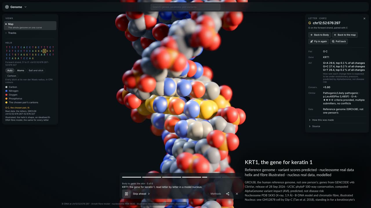
Skin to gene, and back. The Body 2.0 tour's second route in the live app: from upper-arm skin into its outer layer and a skin cell, then to a keratin structure and KRT1, the gene for its K1 component, and back.
MethodsBodyParts3D (Mitsuhashi 2009), TARO (Nagaoka 2004), Z-Anatomy (Kervyn). Skin block/cell illustrated after OpenStax (Betts 2022). K1/K10 coil-1B X-ray structure: 6EC0 (Eldirany 2019). KRT1: GRCh38/GENCODE (Frankish 2023), ClinVar (Landrum 2018), phyloP (Pollard 2010); predicted AlphaGenome variant scores (Cheng 2026; Avsec 2026). Nucleosome: 1KX5 (Davey 2002); Dip-C nucleus (Tan 2018). · real data · simulated · illustrated
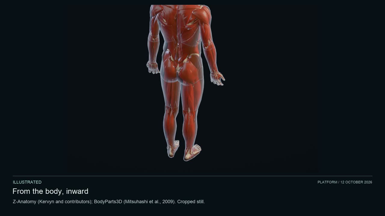
From the body to MYH7, and back. Follow the public walk through a beating heart, illustrated muscle, mouse heart microscopy and a gene for heart muscle, then return to the body. The stops use different references, not one person's anatomy. Microscopy is mouse tissue; GM12878 Dip-C stands in for a cardiomyocyte nucleus. The muscle block is illustrated. Opening and return are cropped still holds with the head out of frame; intervening clips keep their source speed.
MethodsPublic heart-to-gene walk, 12 October 2026. Sources: Z-Anatomy (Kervyn et al.), BodyParts3D (Mitsuhashi 2009), HRA heart (Browne and Schlehlein 2024), Fenton–Karma (1998), Durrer (1970), OpenStax, mouse tomography (Hoshijima 2004; Hayashi 2009), GRCh38 and GM12878 Dip-C (Tan 2018). Contraction, valves and tracers are illustrated, timed by simulated beats. Full Methods names each source and model. Full Methods · real data · simulated · illustrated
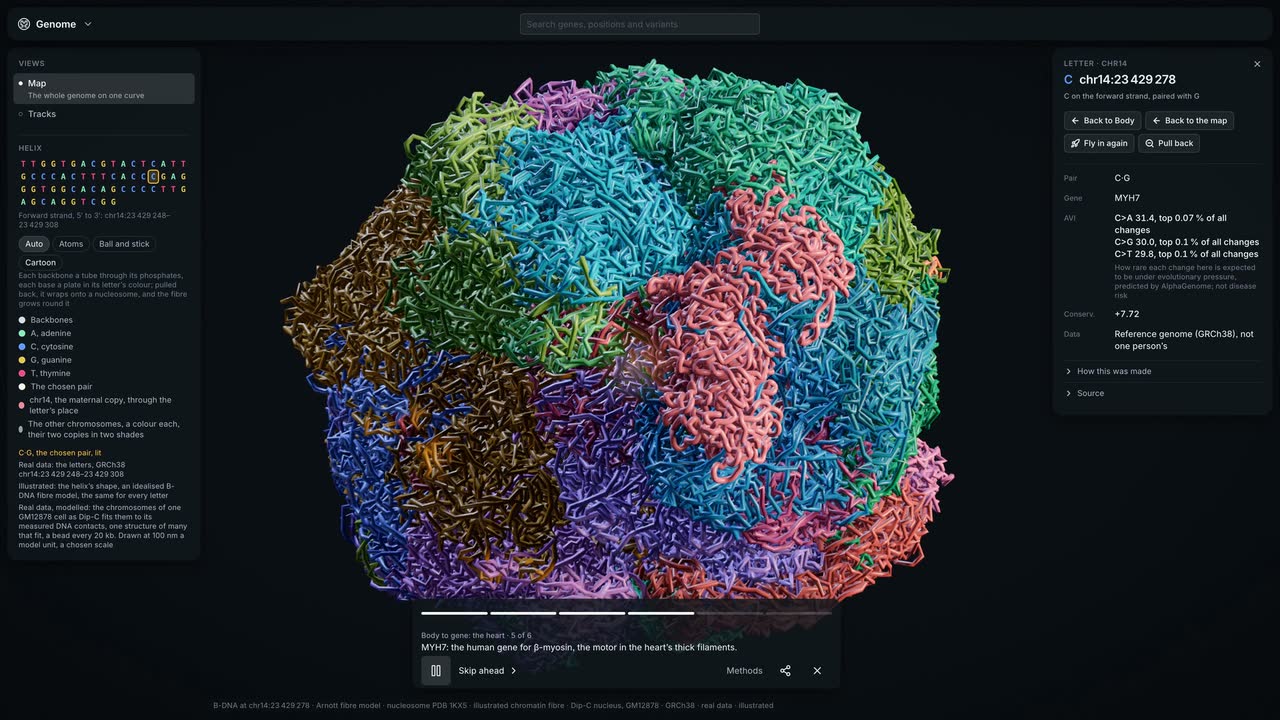
Body to gene, and back. The Body 2.0 tour in the live app: from the whole body into the beating heart, down a heart muscle cell to MYH7, the gene for its motor, and back.
MethodsBodyParts3D, © The Database Center for Life Science (DBCLS), CC BY-SA 2.1 Japan, built on the TARO model from one volunteer's MRI (Nagaoka et al. 2004); reworked by Z-Anatomy (Gauthier Kervyn), CC BY-SA 4.0. With HuBMAP's heart on a simulated heartbeat, mouse heart muscle by electron tomography (CCDB:3611) and MYH7 on GRCh38 with ClinVar, AlphaGenome's predictions, PDB 1KX5's nucleosome and a Dip-C nucleus. This film is shared under the same licence, CC BY-SA 4.0. · real data · simulated · illustrated
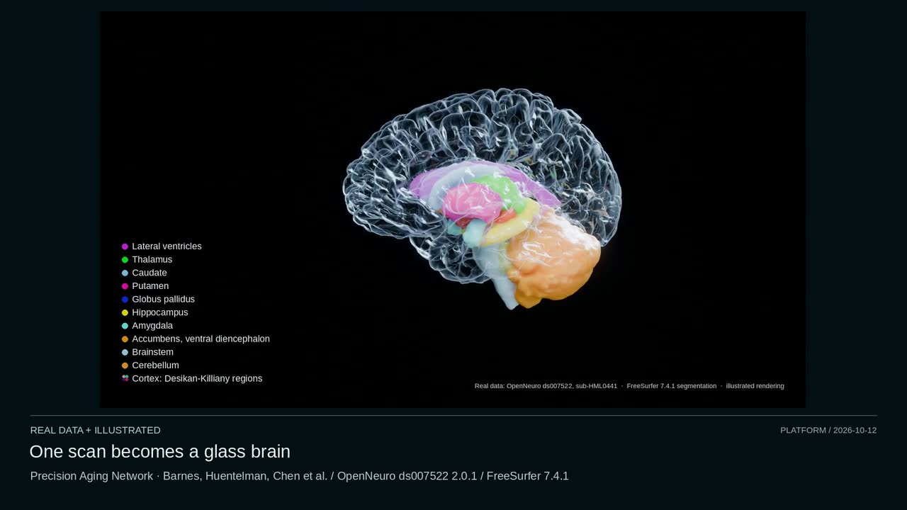
The platform, today · 12 October. From one brain scan to a dividing cell and ribosomes in their own tomogram. Ten separate studies: a water-control focus returns, while running, arterial flow and EEG still expose limits. Corrected 13 October: the faulty glioma-growth excerpt is removed. Separate studies with different clocks and baselines. The larger running model misses its learning check; all six arterial cases still miss; no disjoint EEG range clears all three baselines. Under a stricter check the ribosome-pair count is 8 of 14 additional tomograms. The glioma recurrence view is not fitted to a patient.
MethodsEleven public sources; full models, measurements and credits in Full Methods. Source credit: MNI152 (Fonov 2011), HCP1065 (Yeh 2022), ADAN56 (Boileau 2015), EEG (Wakeman & Henson 2015), ITRUSST (Aubry 2022), running (Carter 2024; Werling 2024), room reflections (Allen & Berkley 1979), cell chromosomes (Thornburg 2026), JCVI-syn3A (CZ CryoET 10442), IXI165 and Precision Aging Network (Barnes, Huentelman, Chen et al.). Full Methods · real data · simulated · illustrated
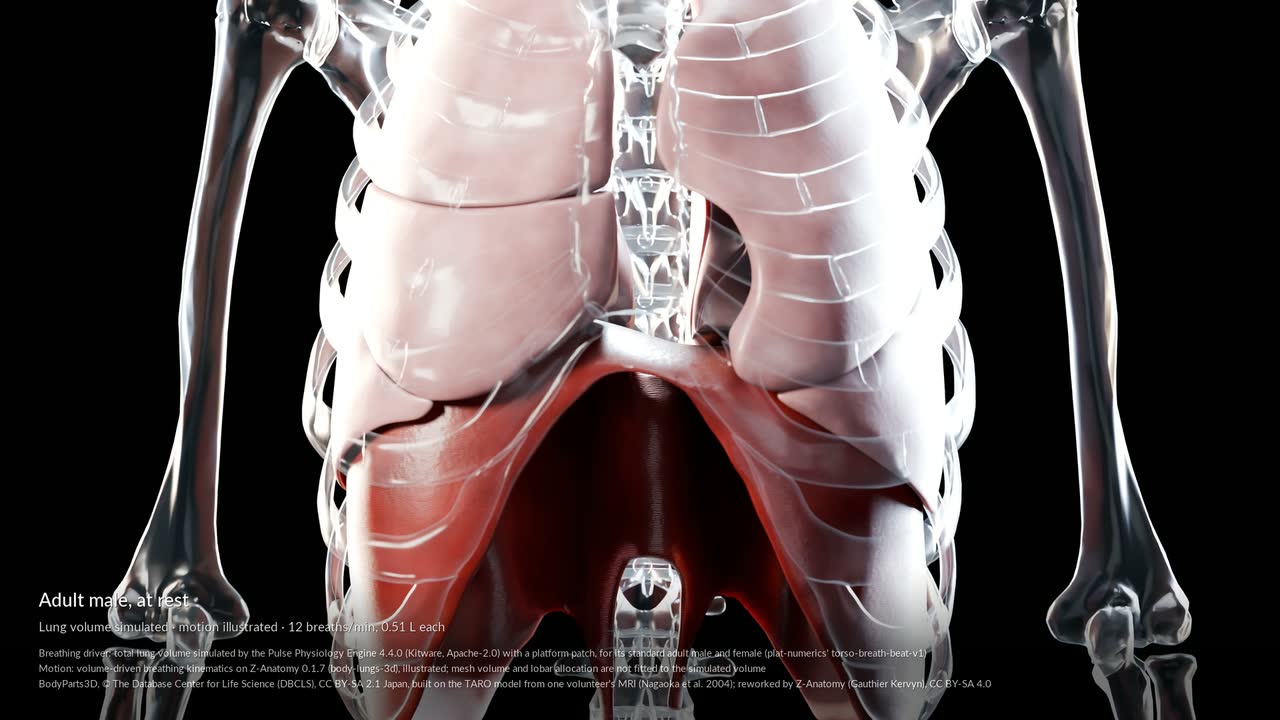
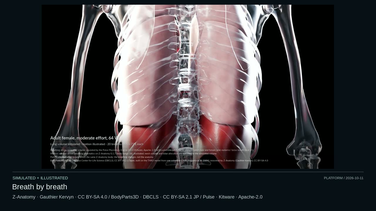
Breath by breath. The Body breathing on simulated lung volumes: Pulse's standard adult male and female at rest, at moderate effort and at vigorous effort, each breath lifting the ribs and lowering the diaphragm.
MethodsBodyParts3D, © The Database Center for Life Science (DBCLS), CC BY-SA 2.1 Japan, built on the TARO model from one volunteer's MRI (Nagaoka et al. 2004); reworked by Z-Anatomy (Gauthier Kervyn), CC BY-SA 4.0. Z-Anatomy 0.1.7 breathing, illustrated, driven by total lung volume simulated in Pulse 4.4.0 (Kitware) for its standard adult male and female at rest, moderate and vigorous effort. This film is shared under the same licence, CC BY-SA 4.0. · simulated · illustrated
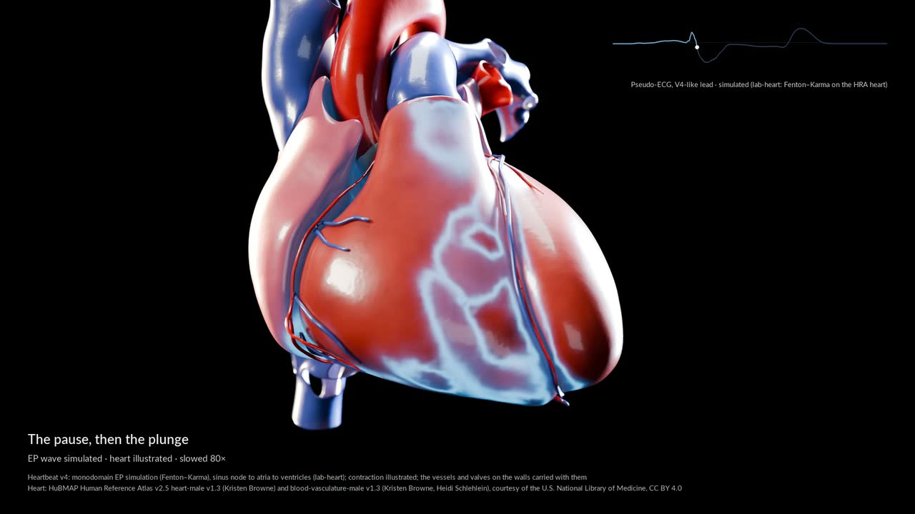
Inside one heartbeat. The Body's heartbeat, one beat slowed up to 80 times: the spark at the sinus node, the pause, the wave through the ventricles, the wringing and the push into the arteries, played on the Body's own anatomy.
MethodsBodyParts3D, © The Database Center for Life Science (DBCLS), CC BY-SA 2.1 Japan, built on the TARO model from one volunteer's MRI (Nagaoka et al. 2004); reworked by Z-Anatomy (Gauthier Kervyn), CC BY-SA 4.0. Heart: HuBMAP Human Reference Atlas, CC BY 4.0. Z-Anatomy 0.1.7; heartbeat v4: monodomain EP, pseudo-ECG and 0D flow simulated; contraction (vessels carried with the walls) and breathing illustrated. This film is shared under the same licence, CC BY-SA 4.0. · simulated · illustrated
Contains flashing or fast-moving patterns that may affect people with photosensitive epilepsy
The platform, today · 11 October. From a breath and a stride to molecules and one base pair. Separate models and measurements, with their own clocks and baselines, cut together; some models still trail simpler predictions. These are separately authored studies and teaching scenes, at different scales and clocks, not one coupled simulation. Some models still trail simple baselines. The Full Methods retain those misses, numerical-only checks and each source’s limits.
MethodsFourteen public sources; full models, limits and credits in the linked Methods. Torso: BodyParts3D, © The Database Center for Life Science (DBCLS), CC BY-SA 2.1 Japan, from TARO (Nagaoka 2004); reworked by Z-Anatomy (Gauthier Kervyn), CC BY-SA 4.0. Pulse: Kitware, Apache-2.0. This film is shared under the same licence, CC BY-SA 4.0. Full Methods · real data · simulated · illustrated
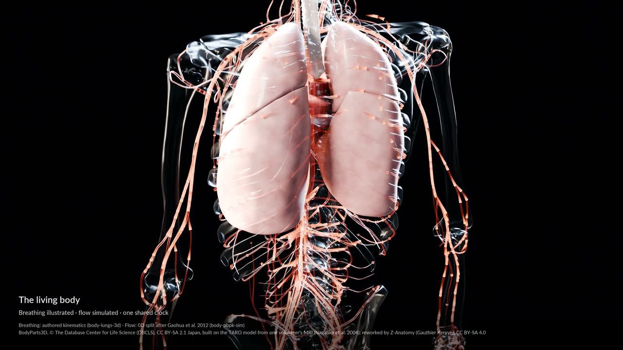
The living body. Breathing, a heartbeat's electrical wave, blood surging through the arteries, a recorded stride and the muscles that drive it: the Body's published tracks, played on its anatomy.
MethodsBodyParts3D (Mitsuhashi et al. 2009) on the TARO model (Nagaoka et al. 2004), reworked by Z-Anatomy (Kervyn); HRA v2.5 heart and vessels (Browne and Schlehlein). Tracks: authored breathing, a simulated monodomain EP wave, a 0D flow split after Gaohua et al. 2012, one Grand Challenge stride (Fregly et al. 2012) shown twice, and OpenSim (Rajagopal et al. 2016) exploratory static optimisation; residuals and saturation in the run header. · real data · simulated · illustrated
Contains flashing or fast-moving patterns that may affect people with photosensitive epilepsy
The platform, today · 10 October. Twelve scenes, each with its own data, model and clock. The remaining glioma tract-spread simulation still allows tumour cells into the brain's fluid spaces. Corrected 13 October: the faulty glioma-growth and mass-effect excerpts are removed. In the remaining tract-spread simulation, tumour cells still enter the brain's fluid spaces, which tumour models normally prevent.
MethodsTwelve published scenes; full models, limits and credits in Full Methods. Source credit: MNI152 / TemplateFlow / McGill; TARO (Nagaoka 2004), BodyParts3D (DBCLS), Z-Anatomy (Kervyn); TractoInferno (Poulin 2022); GC4 (Fregly 2012); Pulse (Kitware); Syn3A (Thornburg 2026); Damagatla 2026; Krokhmal 2025; Wakeman & Henson 2015. Full Methods · real data · simulated · illustrated
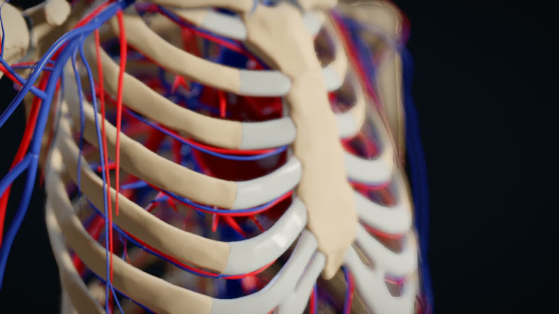
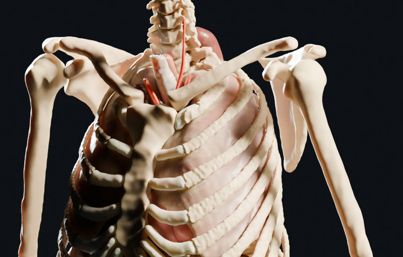
Focus through the ribs. The camera holds on the heart while the focus racks from the front of the rib cage to the back and returns: our engine's depth of field, driven by a camera flight.
MethodsIllustrated: the Z-Anatomy whole-body model (Gauthier Kervyn, from BodyParts3D, DBCLS; CC BY-SA 4.0), from our body-zanatomy 0.1.2 pack, showing the skeleton, vessels and heart. Drawn live by our wgpu engine at 1920×1080, 30 fps: the camera holds on the heart 600 mm away while a scripted flight moves the depth of field's focus from the front ribs (470 mm) to the back ribs (740 mm) and back to the heart. This film is shared under the same licence, CC BY-SA 4.0. · illustrated
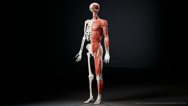
The body in bone and muscle. The whole skeleton, with the muscles of the left half laid over it, from the Z-Anatomy model in our Body 2.0 pack.
MethodsIllustrated: the whole-body model from our body-zanatomy 0.1.5 pack, BodyParts3D, © The Database Center for Life Science (DBCLS), CC BY-SA 2.1 Japan, built on the TARO model from one volunteer's MRI (Nagaoka et al. 2004); reworked by Z-Anatomy (Gauthier Kervyn), CC BY-SA 4.0; face removed by a shear-plane cut. Skeleton on both sides, muscles of the left half. Blender 5.2 Cycles, 512 samples, studio light. This image is shared under the same licence, CC BY-SA 4.0. · illustrated
Heart
The heart, beating as computed.
The reference heart in the donor's chest, its activation and its vessels drawn from the models that produce them.
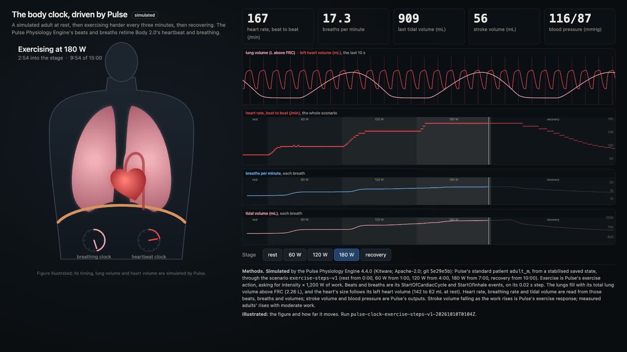
The body clock, driven by Pulse. A simulated adult goes from 71 to 167 heartbeats and from 12 to 17 breaths a minute as the work rises to 180 W, then settles. Body 2.0's heartbeat and breathing follow its beats and breaths.
MethodsPulse Physiology Engine 4.4.0 (Kitware, Apache-2.0), its standard adult male from a stabilised state: rest, then exercise asking 60, 120 and 180 W for three minutes each, then recovery. Beats and breaths are Pulse's cycle events; the lungs and heart follow its lung and left heart volumes. Stroke volume falling with work is Pulse's exercise response; measured adults' rises. Nine real-time seconds per stage; the figure is illustrated. · simulated · illustrated
Contains flashing or fast-moving patterns that may affect people with photosensitive epilepsy
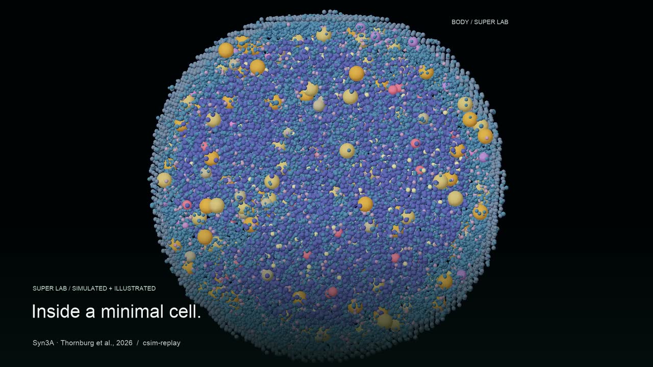
A world within. A 45-second tour from the Body into the cells, currents and living signals of the Super Lab.
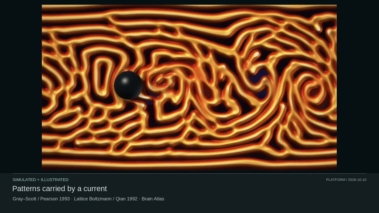
MethodsSynthetic chest and heart: Studio / OpenAI image generation, text only. Syn3A diffusion: Thornburg 2026, Zenodo 15579159 (CC BY 4.0). Turing Current: Gray–Scott chemistry + lattice Boltzmann flow. Shared Lab: Sgro 2015 signaling, Nakajima 2014 chemotaxis and lab-flow. Independent models; authored cameras and rendering. Simulated and illustrated. · simulated · illustrated
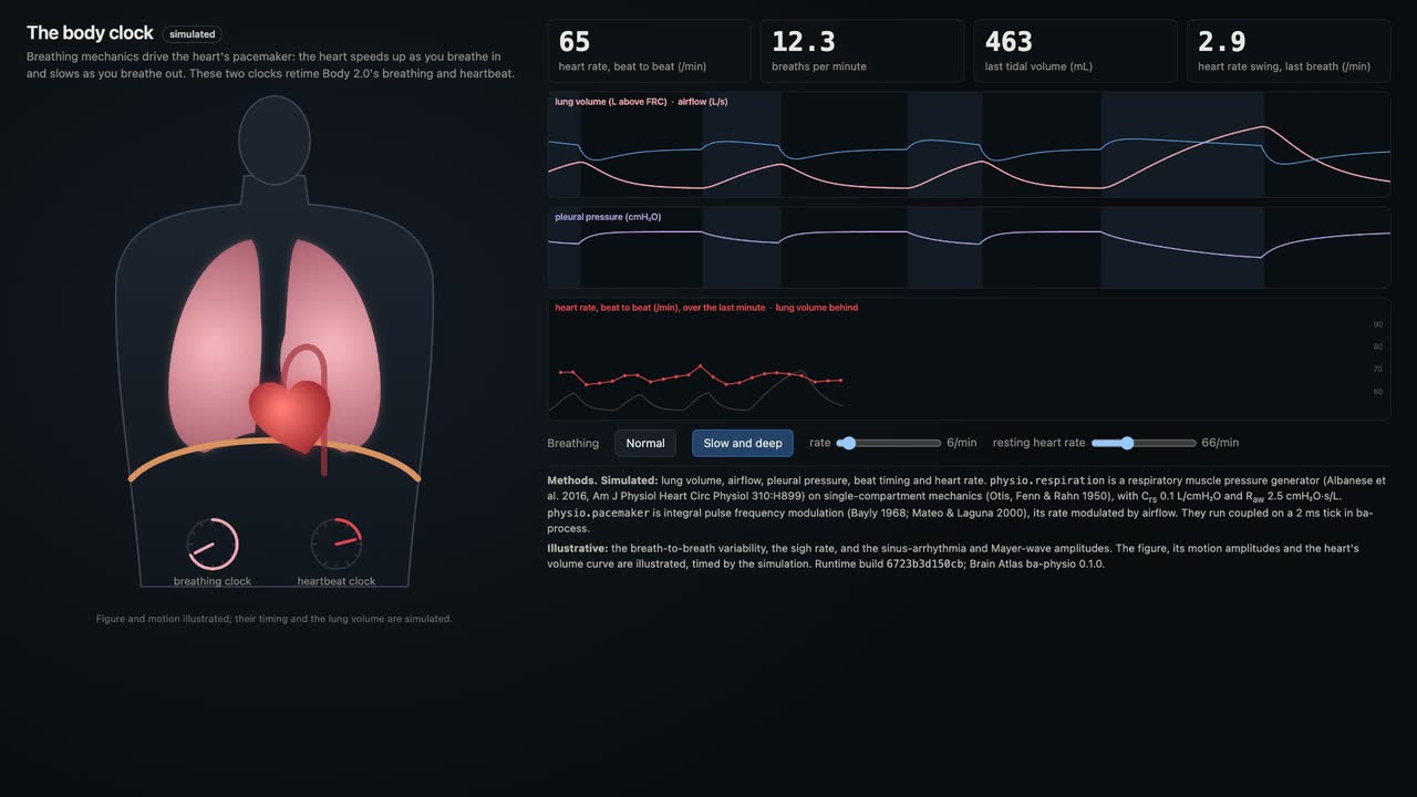
The body clock: breathing paces the heart. Breathe in and the heart speeds up; breathe out and it slows. Switching to six deep breaths a minute widens the swing in each breath from 2.9 to 7.4 beats a minute.
MethodsA respiratory muscle pressure generator (Albanese et al. 2016) on single-compartment lung mechanics (Otis, Fenn & Rahn 1950) drives an integral pulse frequency modulation pacemaker (Bayly 1968; Mateo & Laguna 2000) through airflow, coupled on a 2 ms tick (run body-clock-0.1). There is no baroreflex, so slow breathing swings less than measured. Breath-to-breath variability, the sigh rate and the figure are illustrated. · simulated · illustrated
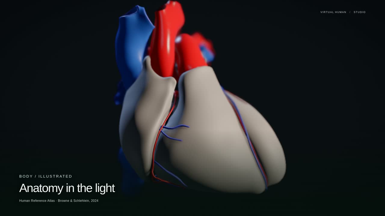
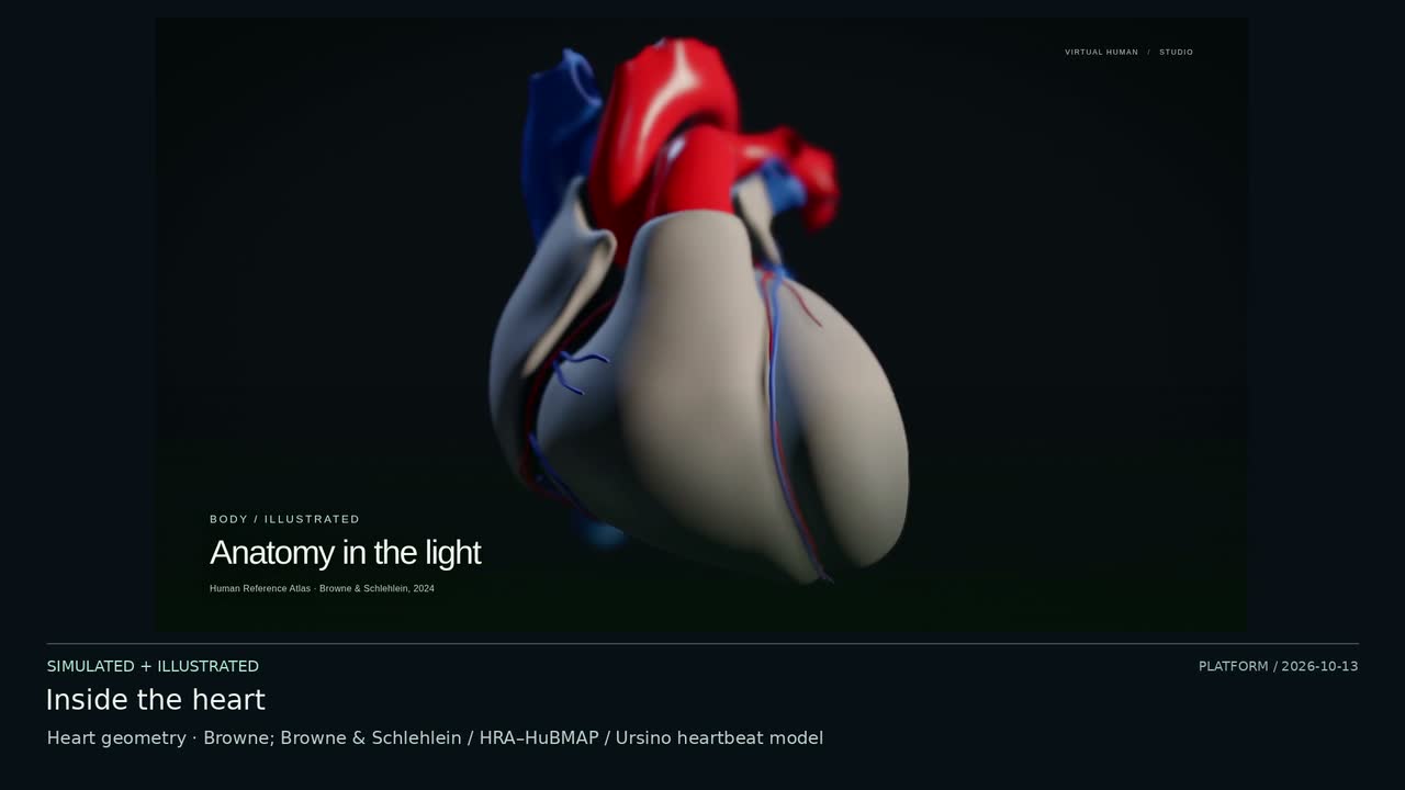
From the heart, inward. A 45-second camera tour from heart anatomy and a recorded heartbeat to a moving protein.
MethodsHRA/HuBMAP heart geometry by Browne and Browne & Schlehlein (CC BY 4.0); an Ursino-based recorded heartbeat at 10× slow motion; PDB 1L2Y by Neidigh et al. (CC0), simulated with AMBER ff14SB/OBC2 at 300 K. Authored lighting and camera paths; the 15 s molecular shot spans 0.449 ps. Simulated, illustrated. · simulated · illustrated
Organs
The organs, where they sit.
Lungs, liver, kidneys and the rest of the viscera, each placed by the fit that put them there.
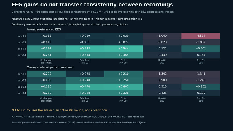
EEG gains do not transfer consistently between recordings. Multipliers fitted on recording 03 improve predictions on recording 05 in 6 of 8 cases. Only 2 of 4 people improve with both EEG preprocessing choices; the pre-written rule requires 3 of 4. Exploratory reuse of four already-seen subjects, with unequal trial counts. The target-fitted bound is not a prediction. Same-trial averages can share artifacts; no fresh validation, source-location or conductivity claim.
MethodsOpenNeuro ds000117, Wakeman & Henson (2015), subjects 01–04. Run-01 MEG-to-EEG statistical maps; one signed multiplier per person and EEG preprocessing choice, fitted on run 03, applied to run 05. Faces-minus-scrambled averages, 0–400 ms; R² relative to zero. Improvement: at least 0.01 above zero, run-01 EEG, run-03 EEG and the unchanged prediction. The run-05 fit uses its target: a best-case bound, not a prediction. · real data · simulated
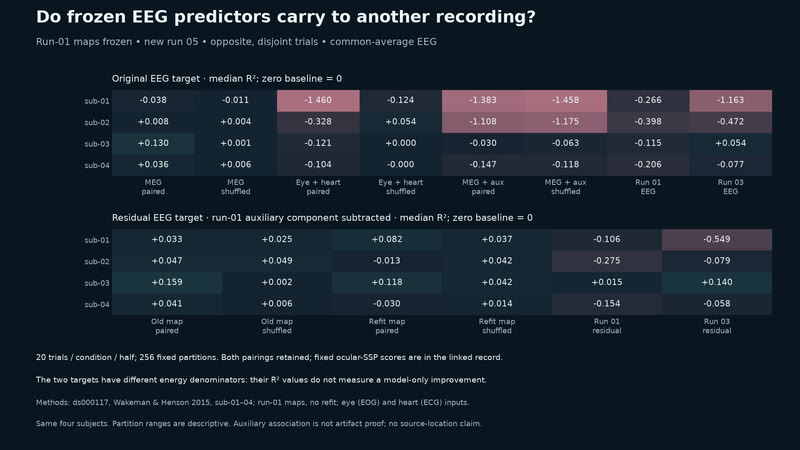
The same-trial pattern partly carries to a new run. Original EEG: MEG sign stability carries in 19/24 same-trial cells, 0/24 disjoint. No disjoint range clears all three baselines. Four reused development subjects, not independent validation. Cells overlap; ranges are descriptive, not confidence intervals. Original and residual R² have different denominators. Auxiliary association can be neural; subtraction may remove it.
MethodsOpenNeuro ds000117 / Wakeman & Henson 2015, sub-01–04 reused. Run-01 MEG/eye/heart maps frozen; run05 faces−scrambled, 0–400 ms. Baselines: zero, run01/run03 EEG. 5/10/20 trials/condition/half; 256 overlapping partitions. Sign: run03 direction in ≥90%. Original/residual R² denominators differ. Descriptive ranges, not confidence intervals or independent validation. Auxiliary subtraction may remove neural signal. · real data · simulated
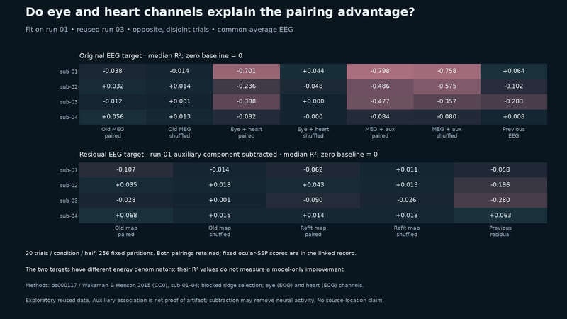
Auxiliary signals share the same-trial gain. Common-average EEG: paired eye/heart maps gain on same trials but lose to zero across disjoint halves. Four reused development subjects; not independent validation. Original and residual targets have different denominators. Partition ranges are not confidence intervals. Auxiliary association does not identify artifact; subtraction and blink projection may remove neural signal.
MethodsOpenNeuro ds000117 2.0.0 / Wakeman & Henson 2015 (CC0), sub-01–04. Statistical MEG/eye/heart ridge maps fit on run 01; reused run 03, faces minus scrambled, 0–400 ms. 256 partitions; 20 trials/condition/half shown. Zero and prior EEG baselines. Original and auxiliary-subtracted targets differ. Exploratory shared preprocessing; ranges are not confidence intervals. Auxiliary association can be neural or artifact; subtraction may remove neural signal. · real data · simulated
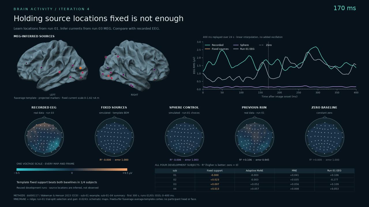
Holding source locations fixed is not enough. The fixed-support template model beats zero and the previous-run EEG in 1/4 subjects. Predictions remain weak; one subject also exposes a numerical instability.
MethodsOpenNeuro ds000117 2.0.0 / Wakeman & Henson 2015 (CC0), sub-01–04, first 300 s of runs 01/03. Run-01 trial splits choose source support, ridge penalty and gain; run-03 MEG supplies currents. Template and sphere EEG predictions share a voltage scale with zero and previous-run baselines. Reused development runs; sub-02 whitening is unstable. FreeSurfer fsaverage average-template cortex; schematic maps. Inferred sources are nonunique. · real data · simulated · illustrated
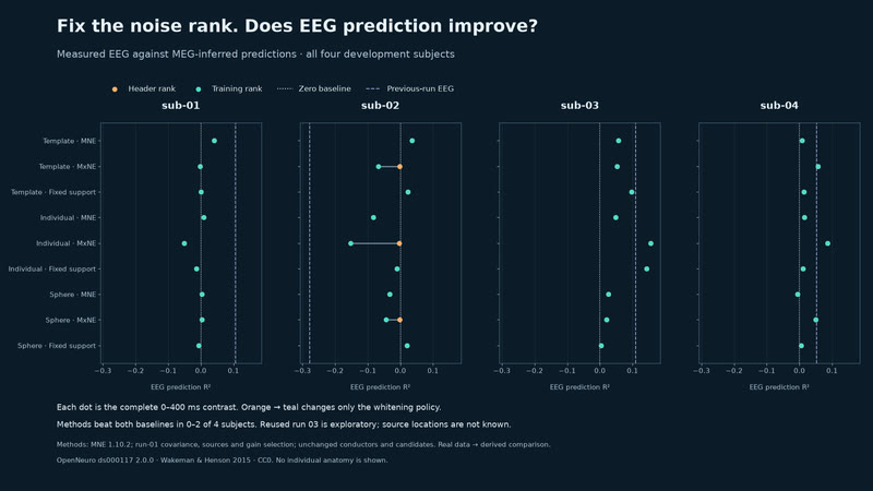
Rank repaired; EEG prediction still misses. The numerical collapse disappears, but sub-02’s sparse predictions worsen. Across four development subjects, methods beat zero and previous-run EEG in 0–2/4.
MethodsOpenNeuro ds000117 2.0.0 / Wakeman & Henson 2015 (CC0), sub-01–04. Run-01 covariance rank is fixed for MNE, MxNE, ridge and residuals; run 03 supplies MEG and measured EEG. Conductors, sources, gain choices and the 0–400 ms window are unchanged. All 128 legacy scores reproduce. Reused run 03 is exploratory. Aggregate plot only; no head or MRI surface. · real data · simulated
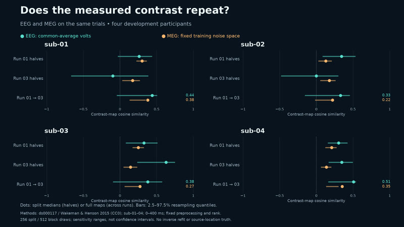
How repeatable is the EEG target? Run-03 EEG split medians range from −0.10 to 0.63 across four development subjects. MEG agreement is modest; the earlier prediction miss remains.
MethodsOpenNeuro ds000117 2.0.0 / Wakeman & Henson 2015 (CC0), sub-01–04. Faces minus scrambled; first 300 s of runs 01/03; 0–400 ms. Common-average EEG; MEG in fixed run-01 noise space. Dots: split medians or full maps. Bars: 2.5–97.5% ranges from 256 splits or 512 ten-trial block draws. Exploratory sensitivity; preprocessing fixed. Aggregate plot only. · real data
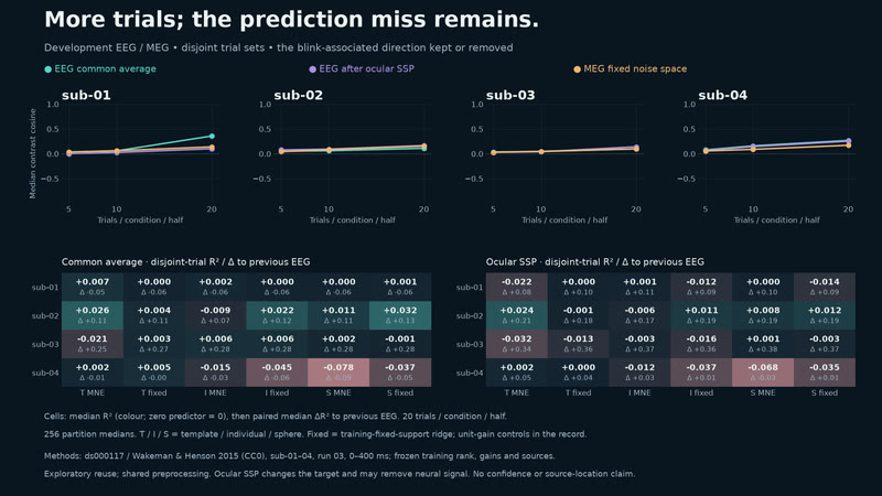
More trials; the prediction miss remains. Median agreement rises with more trials; no frozen model beats both baselines by paired median in all four subjects. Exploratory reuse of four subjects, not independent validation. Partition ranges are not confidence intervals. Disjoint trials share preprocessing; the blink projection changes the target and may remove neural signal.
MethodsOpenNeuro ds000117 2.0.0 / Wakeman & Henson 2015 (CC0), sub-01–04. Faces minus scrambled, run 03, 0–400 ms. Frozen run-01 gains and MEG noise space; template/individual/sphere × MNE/fixed-support ridge. 256 nested disjoint partitions, 5/10/20 trials per condition per half; zero and previous-run EEG baselines. Common-average EEG versus fixed blink projection. Descriptive partition sensitivity; shared preprocessing. Aggregate plot only. · real data · simulated
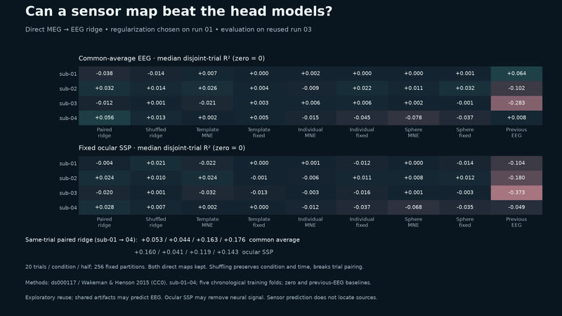
Sensor prediction depends on trial pairing. Same-trial gains shrink across disjoint trials; every disjoint direct-map R² partition range crosses zero. Four reused development subjects, shared preprocessing; not independent validation. Partition ranges are not confidence intervals. Pairing effects may reflect neural signal or artifacts. Blink projection may remove neural signal; paired and shuffled fits use separately tuned shrinkage.
MethodsOpenNeuro ds000117 2.0.0 / Wakeman & Henson 2015 (CC0), sub-01–04. MEG→EEG ridge; paired/shuffled fits tuned separately on run 01; reused run 03, faces minus scrambled, 0–400 ms. 256 same/disjoint partitions; 20 trials/condition/half shown. Zero and prior EEG baselines. Exploratory shared preprocessing; ranges are not confidence intervals. Pairing can reflect neural signal or artifacts; blink projection may remove neural signal. · real data · simulated
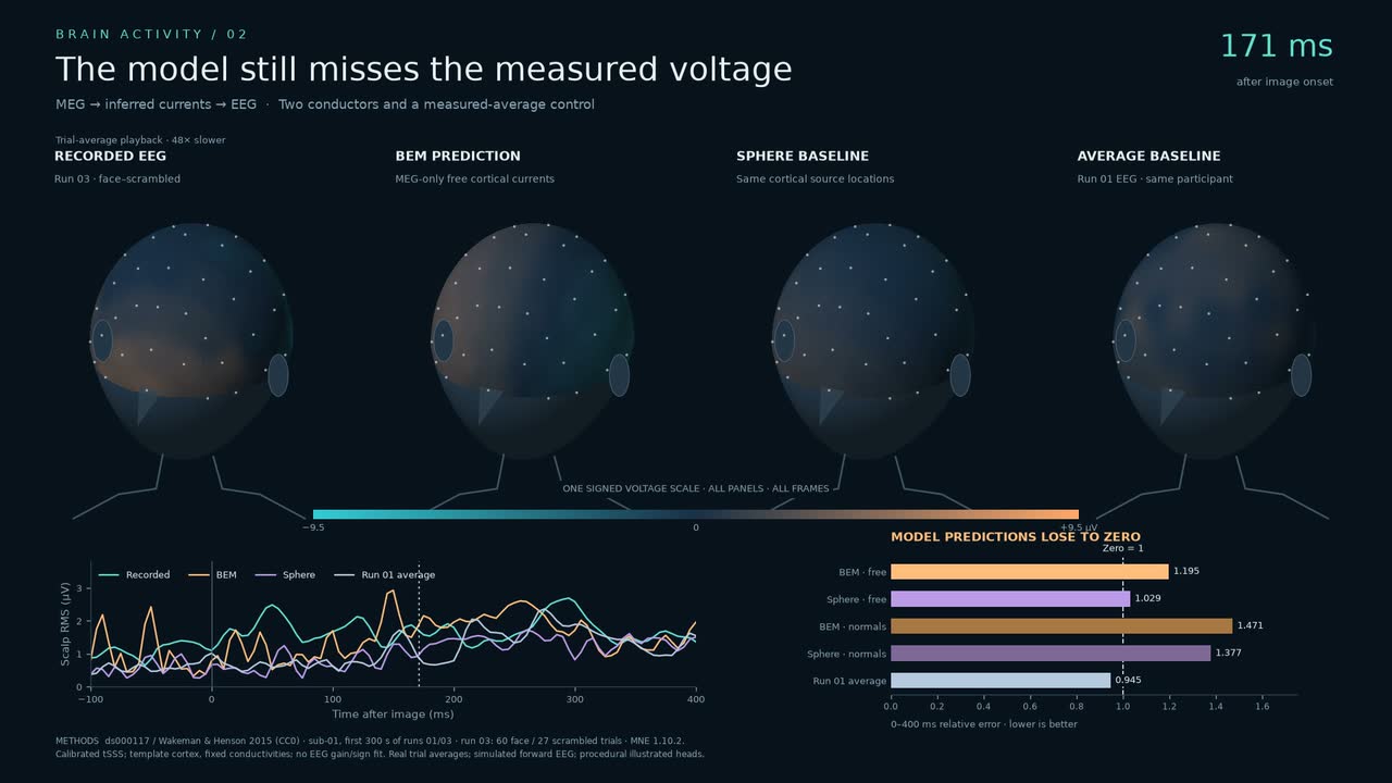
Measured EEG challenges the model. The head models lose to zero. The previous-run average does better.
MethodsModel predictions lose to zero: BEM error 1.195, sphere 1.029; run-01 EEG average 0.945 (zero 1). OpenNeuro ds000117 / Wakeman & Henson 2015, CC0, sub-01 runs 01/03, first 300 s. MNE physical currents on template cortex; fixed conductivities, no EEG gain/sign fit. Common-average face–scrambled EEG; same trials and signed voltage scale. Procedural head. · real data · simulated · illustrated
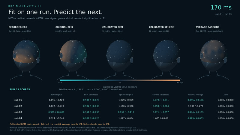
Fit on one run. Predict the next. Calibrated BEM beats zero in 4/4 recordings, but the previous-run EEG average in only 1/4.
MethodsOpenNeuro ds000117 2.0.0 / Wakeman & Henson 2015 (CC0), sub-01–04, first 300 s of runs 01/03. MNE on template cortex; tSSS and common-average face–scrambled EEG. A signed scalar gain and skull conductivity are fitted on run 01, then fixed for run 03. Original models, zero and same-subject run-01 EEG are controls. Exploratory transfer, not conductivity identification. Sub-01 at 170 ms; one voltage scale; procedural heads. · real data · simulated · illustrated
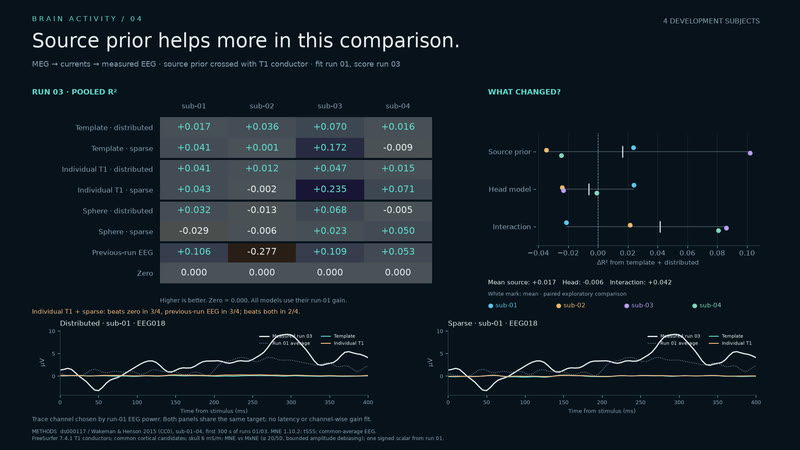
Source prior helps more in this comparison. Across four development subjects, source-prior change adds 0.017 mean R²; head-only change −0.006. The combined model beats both baselines in 2/4.
MethodsOpenNeuro ds000117 2.0.0 / Wakeman & Henson 2015 (CC0), sub-01–04, first 300 s of runs 01/03. MEG-only source fits with distributed MNE and sparse MxNE, crossed with template, individual T1 and sphere conductors. Gain and sparse alpha are chosen on run 01, then fixed for run 03 EEG scoring. Zero and previous-run EEG are baselines. Same candidate currents and fixed skull conductivity. Four explored subjects; aggregate plots only. · real data · simulated
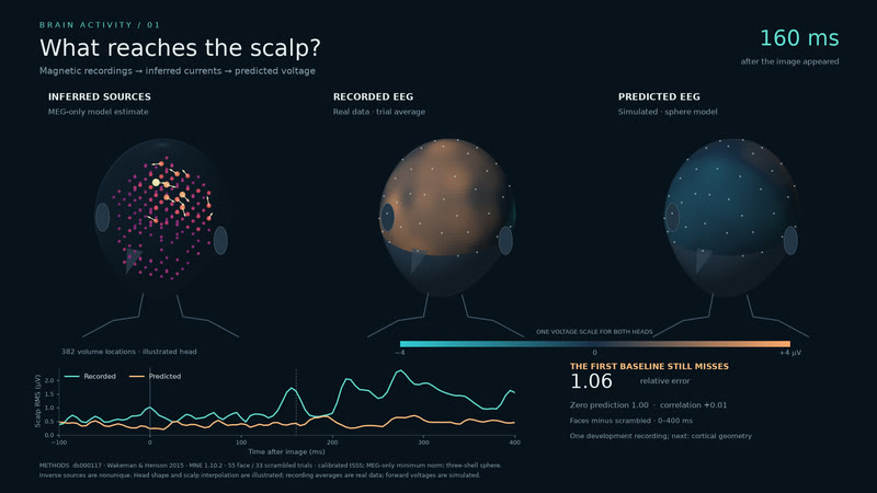
Brain activity, measured and predicted. MEG estimates inside the head, with recorded and predicted EEG side by side.
MethodsDoes not beat zero: EEG prediction error 1.055 versus 1.000. MEG-derived MNE currents drive a three-shell sphere; recorded EEG is the comparison. OpenNeuro ds000117 / Wakeman & Henson 2015, CC0, sub-01 run 01, face–scrambled trial average. Generic illustrated head. Fixed ±6 µV, 72× slow motion; no gain or polarity fit. · real data · simulated · illustrated
Muscles & joints
What moves the skeleton.
Muscles and joints as the data gives them, and motion where a model supplies it.
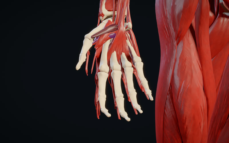
Opening a hand. A hand-opening motion retargeted onto the atlas, with model-estimated muscle activation.
MethodsARMS Lab, McFarland et al. 2023: averaged letter-O motion from two participants, 1–8.995 s. Activations recomputed by static optimisation of 43 muscles, with substantial artificial torque assistance at the ring finger’s middle joint. Whole flexor pollicis brevis shown on its superficial head; palmar interossei averaged by maximum isometric force. Rigid motion and left mirror illustrated. Z-Anatomy (Kervyn), CC BY-SA 4.0, from BodyParts3D (DBCLS). · real data · simulated · illustrated
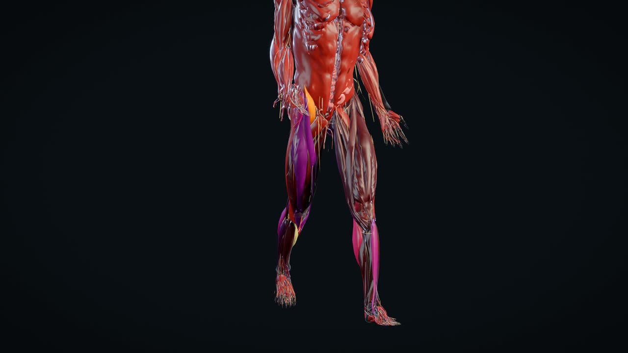
A step, with muscle and nerve cues. A recorded step, replayed slowly with model-estimated muscle activation and illustrated nerve cues.
MethodsWalk: OpenSim’s Rajagopal example, motion_capture_walk.trc (opensim-models d9b05d47), 0.45–1.65 s, retargeted onto illustrated anatomy. Activation: static optimisation with the Rajagopal et al. 2016 model in OpenSim. Nerve cues: drawn routes timed to activation. Anatomy: Z-Anatomy (Gauthier Kervyn), CC BY-SA 4.0, from BodyParts3D (DBCLS). External genital structures are hidden in this view. · real data · simulated · illustrated
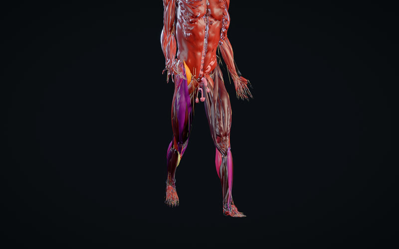
A step, with muscle and nerve cues. A recorded step, with muscle activation estimated by a model and drawn nerve cues timed to that activation.
MethodsWalk: OpenSim’s Rajagopal example, motion_capture_walk.trc (opensim-models d9b05d47), 0.45–1.65 s, retargeted onto illustrated anatomy. Activation: static optimisation with the Rajagopal et al. 2016 model in OpenSim. Nerve cues: drawn routes timed to activation. Anatomy: Z-Anatomy (Gauthier Kervyn), CC BY-SA 4.0, from BodyParts3D (DBCLS). · real data · simulated · illustrated
Skin
A synthetic skin.
Portrait studies on a generic synthetic adult: procedural skin, hair and lip materials, lit and rendered in Blender. Illustrated; not a real person.
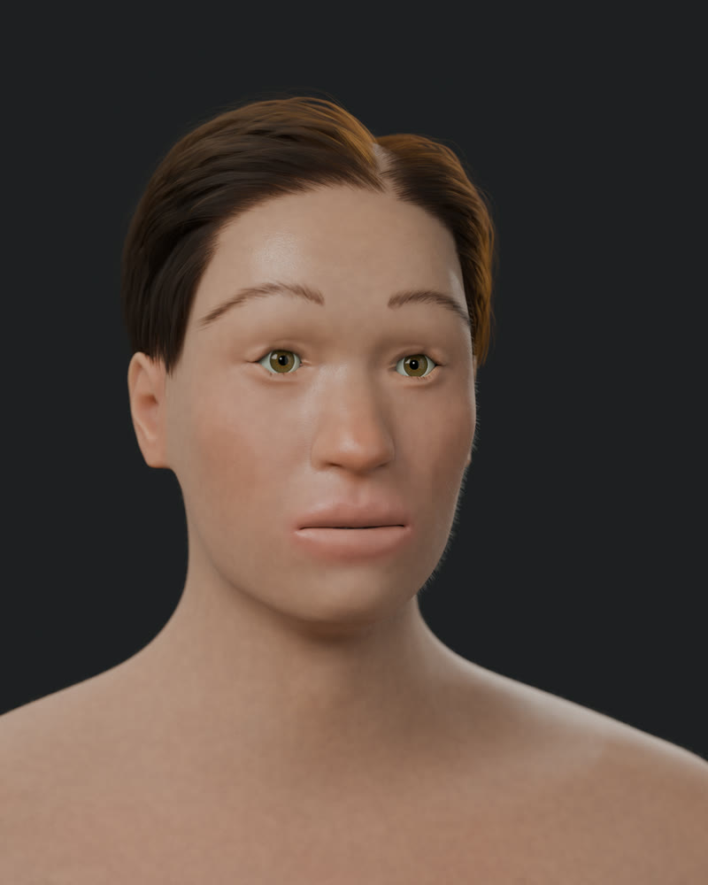
Synthetic skin — lip material study. A procedural adult portrait with fine skin texture and soft lip highlights. Illustrated (synthetic human; not a real person).
MethodsBlender 5.2.2 Cycles OptiX; MPFB2 generic adult configured at age 35 with procedural skin and lip materials; no real person's likeness and no scan fitting. Matched 1600×2000 portraits at 512 samples on one NVIDIA L40S; this is the lip-material variant (B). · illustrated
Cells & molecules
Down to the cell.
From an organ to its tissue, its cells and their molecules: each step from a measured dataset or a model that names it.
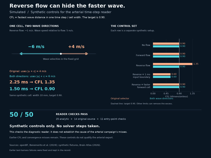
Reverse flow can hide the faster wave. In a synthetic reverse-flow cell, the original time-step selector gives CFL 1.35 against 0.90; using both wave directions gives 0.90. All 50 reader checks pass. This figure shows synthetic fixtures. Synthetic controls only: this figure does not show retained arterial states. Earlier campaign CFL and convergence misses remain; these checks do not qualify the arterial export.
MethodsSources: openBF, Benemerito et al. (2024); synthetic fixtures, Brain Atlas (2026). Cell width 10 mm; wave speed 5 m/s; target CFL 0.90. Analytic checks 25/25, original-source integration 14/14, reader entry-point checks 11/11. Two earlier harness failures were fixed and retained. For these controls, no campaign states were read or solver steps taken. · simulated
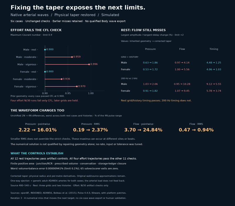
Fixing the taper exposes the next limits. Rest grid/history timing now passes, but flow-amplitude changes reach 4.14% (male) and 5.56% (female). At 200 Hz, flow changes reach 10.28% and timing 5.55%; male pressure also misses at 2.06%. Effort CFL reaches 0.996 against 0.9. Conservation, finite area, junction/RCR, prescribed volume and ledger controls pass on all 16 completed trajectories. All six cases miss: four effort cases exceed CFL 0.9; their later grids are held. Both rest cases miss flow amplitude and 200 Hz sampling; male rest also misses sampled pressure. Same 2% comparison limits, no qualified export. One-way coupling; generic adult geometry for both sexes.
MethodsSources: openBF, INSIGNEO; ADAN56, Boileau et al. (2015); Pulse 4.4.0, Kitware, with platform patches. Corrected physical taper, unchanged T adapter and inputs, source 480–540 s. Complete prior campaign retained. Rest: N/2N/4N, 30/60 s histories; effort: N/30, later grids held after CFL misses. Same strict 2% limits; both arrival detectors kept. One-way generic geometry; no qualified export. · simulated
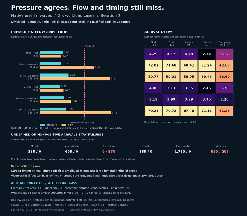
Pressure agrees. Flow and timing still miss. Pressure-amplitude changes stay below 1.30%; flow reaches 3.18% and finite arrival-delay change 72.02%, against unchanged 2% limits. All 36 runs pass conservation, finite-area, CFL, junction/RCR, prescribed-volume and ledger controls. Carotid timing and invalid vigorous tibial arrivals remain iteration targets. Inherited openBF taper code loses over 99.9% of the specified radius change in 39 segments at N; refinement loses more. A corrected-taper run is planned. Pressure amplitudes pass everywhere. Flow amplitudes miss in male moderate, male vigorous and female vigorous; every case misses timing. Anterior-tibial foot rows are invalid in both vigorous cases. All six remain MISS; no qualified Body wave export. Inherited openBF taper code loses over 99.9% of the specified radius change in 39 segments at N; refinement loses more. A corrected-taper run is planned.
MethodsopenBF, INSIGNEO; ADAN56, Boileau et al. (2015); Pulse 4.4.0, Kitware, with platform patches. Native ejection at source 480–540 s, one-way coupling. Six cases, three grids/two histories; unchanged 2% limits; Chiu (1991) tangent beside the original 10% foot. No qualified export. Inherited taper code loses over 99.9% of 39 specified radius changes at N; the corrected arm is planned. · simulated
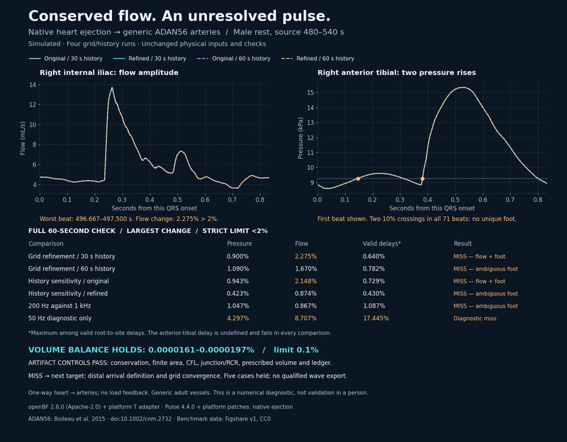
Conserved flow. An unresolved pulse. A miss moves the iteration target: anterior_tibial_R has a double upstroke; one internal_iliac_R beat changes 2.28% under refinement (limit 2%). Artifact controls pass: conservation, finite area, CFL, junction/RCR, prescribed volume and ledger. Inherited openBF taper code loses over 99.9% of the specified radius change in 39 segments at N; refinement loses more. A corrected-taper run is planned. anterior_tibial_R has a double upstroke in all 71 beats; one internal_iliac_R beat changes 2.28% under refinement (limit 2%). This miss moves the iteration target. First campaign held five cases; no qualified wave export. Generic arteries; one-way coupling. Historical female 240 W pressure miss remains open. Inherited openBF taper code loses over 99.9% of the specified radius change in 39 segments at N; refinement loses more. A corrected-taper run is planned.
MethodsopenBF, INSIGNEO; ADAN56, Boileau et al. (2015); Pulse 4.4.0, Kitware, with platform patches. Native ejection at source 480–540 s, one-way coupling. Male rest, two grids/two histories, unchanged 2% rule. First-case MISS; no qualified export. Inherited taper code loses over 99.9% of 39 specified radius changes at N; the corrected arm is planned. · simulated
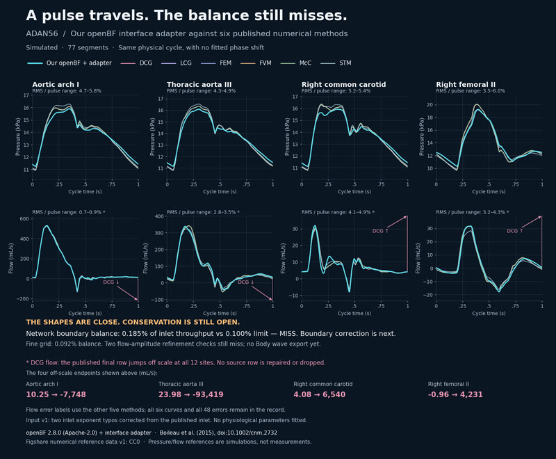
A pulse travels. The balance still misses. Pressure shapes agree within 3–6% of the published pulse range (RMS); flow within 0.7–4.9%, apart from DCG’s anomalous final row. Network boundary balance still misses: 0.185% vs 0.100%. The boundary correction is next.
MethodsopenBF 2.8.0 (Apache-2.0) with our interface adapter, generic ADAN56, simulated. Boileau et al. (2015), doi:10.1002/cnm.2732; six numerical methods, Figshare v1 (CC0). Four mid-vessel sites, cycle 9–10 s; no fitted shift. Two inlet exponent typos repaired as input v1. The DCG flow endpoint is retained off scale. Pressure agreement is RMS error/pulse range; conservation remains unresolved. · simulated
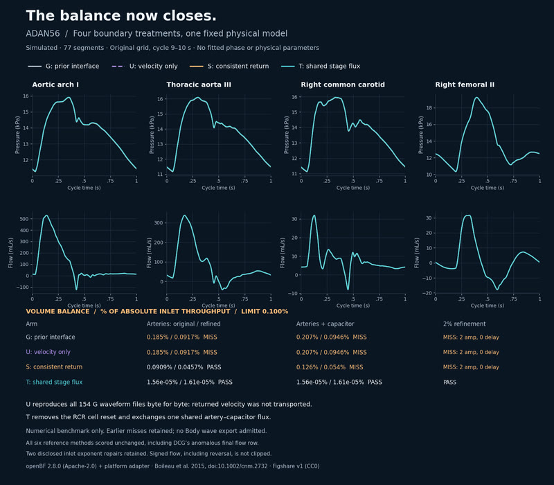
The balance now closes. G’s artery balance misses at 0.185% on the original grid (0.092% refined); T gives 1.56e-05%/1.61e-05%, against a 0.100% limit. U, S and T separate returned state from shared boundary exchange. Refinement results and remaining caveats are shown. Numerical benchmark; no Body wave export is admitted. T passes the numerical gates shown. All earlier misses and the stopped harness attempt remain recorded. DCG’s last-row anomaly and the two disclosed inlet repairs are unchanged.
MethodsopenBF 2.8.0 (Apache-2.0) + platform boundary adapter; generic ADAN56, simulated. G/U/S/T share the same inlet, wall law and RCR parameters. Four mid-vessel sites, cycle 9–10 s; no fitted shift. Boileau et al. (2015), doi:10.1002/cnm.2732, six numerical methods from Figshare v1 (CC0). Two inlet exponent repairs and the DCG endpoint anomaly remain disclosed. No Body wave export is admitted. · simulated
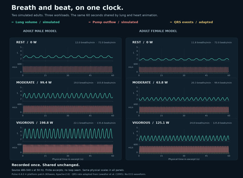
Breath and beat, on one clock. Six recordings connect lung and heart animation through shared physical time. The data is public and consumer checks passed; live animation is a separate rollout. The VO2, kidney/skin and historical pressure misses remain.
MethodsPulse 4.4.0 + platform patch (Kitware, Apache-2.0): native lung volume and left-heart outflow. QRS timing adapted from Lewalter et al. (1995), with a 72/min model intercept and lab-heart's activation-onset convention; no ECG waveform. Six 50 Hz excerpts from source 480–540 s, with identical physical scales. Numerical consumer checks passed; app animation is a separate rollout. Pressure floor: model rest MAP minus 10 mmHg, not measured. · simulated
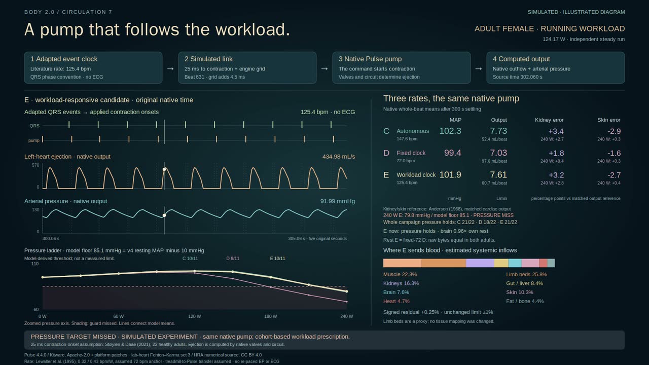
A pump that follows the workload. Workload-driven beats restore three pressure cases. One still misses; kidney and skin errors remain. Floor: v4 model's own resting MAP minus 10 mmHg (model-derived threshold, not a measured limit).
MethodsPulse 4.4.0 + platform patch (Kitware, Apache-2.0). Lewalter 1995 sex-specific cohort work slopes pace an adapted QRS clock; 72 bpm rest and treadmill-work transfer assumed, no ECG. Phase convention: lab-heart Fenton–Karma/HRA (CC BY 4.0). Assumed 25 ms contraction delay: Støylen & Daae 2021; 20 ms grid, native valves. Pressure 21/22; female 240 W 79.8<85.1 mmHg. Kidney/skin errors: Anderson 1968. Brain Atlas SVG. · simulated · illustrated
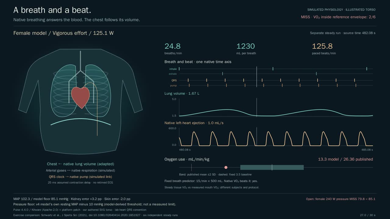
A breath and a beat. Breathing follows workload, but oxygen use misses all four exercise references. The earlier pressure miss and kidney/skin errors remain. Floor: v4 model's own resting MAP minus 10 mmHg (model-derived threshold, not a measured limit).
MethodsPulse 4.4.0 + platform patch (Kitware, Apache-2.0), native lungs and blood gases. Tissue VO2 vs mouth VO2: Schwartz et al.2021; independent steady runs, not matched subjects. Workload-paced QRS convention: lab-heart (Fenton–Karma/HRA, CC BY 4.0); no retimed ECG. Lewalter1995 rate slope; assumed25ms delay, Støylen & Daae2021. Our SVG chest is adapted from lung volume. · simulated · illustrated
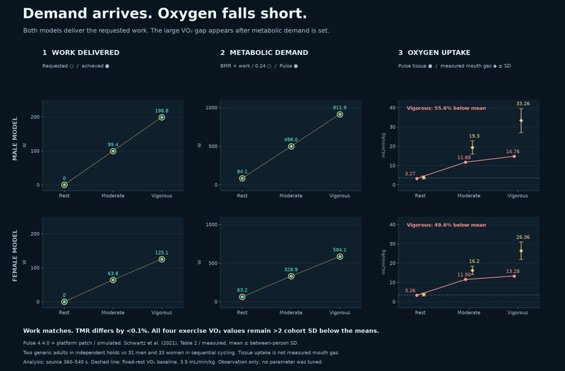
Demand arrives. Oxygen falls short. The models report the requested work and expected metabolic demand, yet vigorous oxygen uptake is 55.6% and 49.6% below the measured cohort means. The shortfall is downstream; its mechanism is not yet separated. Kidney/skin errors and the historical model-pressure miss remain.
MethodsPulse 4.4.0 + platform patch (Kitware, Apache-2.0), simulated; unchanged six-case results, means over 360–540 s. Expected TMR = basal demand + work/0.24. Schwartz et al. (2021), Table 2: measured mouth-gas VO2, cohort mean ± SD. Two generic models in independent holds are not matched to the 31 men and 33 women in sequential cycling. Dashed VO2 baseline: fixed 3.5 mL/min/kg. No tuning. · real data · simulated
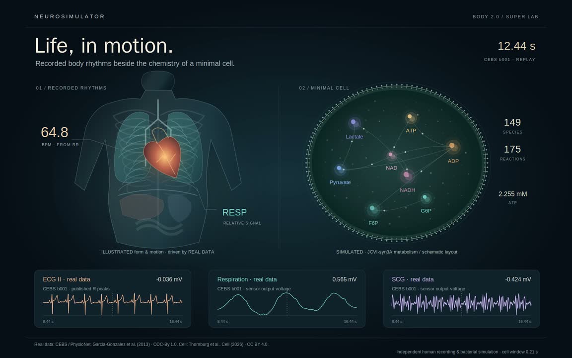
Life, in motion · measured mechanics. Electrical beats, chest vibration and breathing from one recording, beside simulated minimal-cell chemistry.
MethodsCEBS b001 / PhysioNet (Garcia-Gonzalez 2013; Goldberger 2000), ODC-By 1.0: simultaneous ECG II, respiration and SCG, 60–100 s. Display at 500 Hz; R peaks retain 5 kHz timing. RESP/SCG are sensor voltages. Body motion is illustrated; no inhale polarity inferred. Independent JCVI-syn3A simulation: Thornburg et al., Cell (2026), CC BY 4.0, one recorded second. Real data, simulated, illustrated. · real data · simulated · illustrated