Fly into syn3A
From a real cell imaged by cryo-electron tomography, through a model cell, to one gene's letters on a drawn helix.
in development · real data · simulated · illustrated
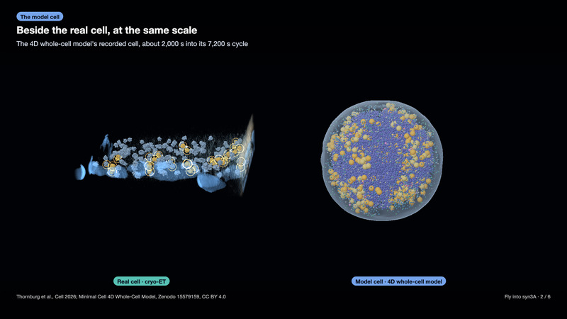
This is a real cell. JCVI-syn3A is a bacterium built in the lab with about 500 genes. One was frozen whole and imaged by cryo-electron tomography. Its ribosomes sit where the tomogram puts them, and 32 of them form 16 pairs, each turned roughly a half turn to its partner, though its genome encodes no known ribosome-pairing factor.
Beside it, at the same scale, is the same organism in the 4D whole-cell model. The flight goes inside the model cell 2,002 s into its cycle, among its ribosomes and along its chromosome, then into another of the model's cells whose chromosomes have just divided between its daughters. It ends on one gene's letters as atoms.
Explore the demo
Needs WebGPU. Live route: LIMIT, with warning, static poster and deliberate start; the film’s PASS applies only to the film.
The result
A real cell beside its model, then a flight from the cell to one gene.
The captured film passes its scene and flash checks. The two earlier captures remain unpublished.
- In this tomogram, 32 ribosomes form 16 pairs 23.9–26.0 nm apart, each turned roughly a half turn to its partner; what holds them together is unknown.
- The flight enters the model cell at 2,002 s. Its chromosome comes from the window run at 1,996 s, which that lattice state holds.
- The model has 558 full ribosomes. The model records their positions, not their orientations; each is drawn here as a sphere.
- The model uses a 10 nm lattice. The re-simulated chromosome represents 10 base pairs with each bead.
- The chromosome is our re-simulation from the model's deposited inputs; the deposit holds no recorded bead positions.
- One gene's letters, on a drawn helix: the first 60 letters of rrsA, the gene for 16S ribosomal RNA, drawn as atoms on an idealised DNA helix.
- The film then enters cell 35, a different model cell, at 5,919 s. Its chromosomes are our re-simulation after partition into the daughters.
The film shows cell 35 as a separate chapter. The live browser route stays with cell 21 and keeps its own photosensitivity warning and deliberate start; the film’s check does not clear the live route.
Checks that failed
- Earlier film: original capture, unpublished
- Luma 6.5/s; linear 7.0/s · The original capture failed the same pinned checker and is superseded by the film linked here.
- Earlier film: first fix, unpublished
- Luma 4.0/s at 39.13 s · The first fix failed the pinned checker and the scene check’s 53 s view. It is not the film linked here.
Checks that passed
- Film scene: sampled views present
- 26 of 26 sampled views passed · The captured scene’s still check passed with no reported page errors, including the separate cell-35 chapter.
- Film photosensitivity check
- Luma 2.5/s; linear 2.0/s; red 0/s · The pinned checker passed the staged film’s exact bytes. Its worst luma window starts at 25.67 s. Its 12 controls passed.
- Changed film windows
- 0.5/s and 2.0/s in luma · The changed 38–42.5 s and 50–56 s ranges remain below the checker’s 3.0/s limit.
Methods
real data · simulated · illustrated
Measured: the cryo-electron tomogram of JCVI-syn3A, CZ CryoET Data Portal 10442, run 24jul17a_Position_2_3. The depositors' denoised AreTomo3 reconstruction (about 0.501 nm voxels) is displayed at about 2 nm per voxel. The membrane and cell masks are MemBrain-seg and SABER predictions, respectively; their resampled forms remain predicted. The depositors' 192 ribosome picks, the refined picks and their pairs are derived. The same cell's 198 template-matched, refined picks include 188 inside the cell mask; each is drawn with the reference ribosome surface EMD-75898 at its refined position and orientation. Its pairs have K 32 against the shuffles' 95th percentiles of 3 and 4. The pairs are a development finding; its confirmatory check (registration C) is in progress; the Lab card's caveat applies. Simulated: Thornburg et al.'s 4D whole-cell model (Cell 2026), cell 21, trajectory MinCell_1, played from 1,940 to 2,002 s of its 7,200 s recording. The lattice snapshot is drawn, never stepped. The model has 558 full ribosomes. The model records their positions, not their orientations; each is drawn here as a sphere. Its DNA sites are repainted from our chromosome re-simulation using the deposit's inputs; the deposit holds no bead positions. Cell 21's chromosome window ran at 1,996 s. The film was captured from source commit ab390a8e4e693ff3d56f139874c451436c425460. The film then shows cell 35, a different model cell, at 5,919 s, from the Brownian-dynamics export csim/bd-chain/cell35-dev-partition-v1, window w05919. Its chromosome source is w05919/play/system.bdsys.gz (SHA-256 5730ee72c7aa1a5d576366bab6b61e61519106e0e4d5c2654d785d998998d386); its gene table is cell35-v1-w05919.genes.json (SHA-256 5d58dbb1eea295e7977a25847c1d6941df28fd473d448d7e99d7410ebc153c34). At partition at 5,827 s, the deposit's unmodified functions wrote the daughters' directives and starting coordinates; its btree_chromo and LAMMPS ran them, and polymer-bd resumes from the joined result. From window 462 at 5,771 s, a protocol correction moves any DNA bead left outside the shell by a neck step or soft minimize to 2 Å inside it, then repeats the deposit's soft minimize. The genes are placed from the deposited GenBank file and lit along the chain. Reference letters on an illustrated helix: rrsA (JCVISYN3A_0069), complement(55713..57247), 1,535 base pairs in the deposited sequence; its first 60 letters are laid out as atoms on Arnott and Hukins' idealised B-DNA. Colours, radii, captions and the camera path are illustrated. The live browser route stays with cell 21 and never loads cell 35's development files. The 75 s film is a recorded flight, not a live simulation.
The pairs are a development finding with confirmation in progress. Cell 35's separate chromosomes come from our development chain.
Sources
- Ali et al. (2024), CZ CryoET Data Portal dataset 10442 · CC0
- Thornburg et al. (2026), Minimal Cell 4D Whole-Cell Model, Zenodo 15579159 · CC BY 4.0
- Hutchings et al., JCVI-syn3A ribosome, EMD-75898 · CC0
- GenBank CP016816.2, as included in the whole-cell model deposit · Public sequence; deposited model data CC BY 4.0
- Arnott and Hukins (1972), idealised B-DNA geometry · Published geometric parameters; no article figures reproduced
Credits
- Ali, Peck, Yu, Schwartz, Glass, Zheng, Paraan and Montabana · The cell's cryo-electron tomography data
- Thornburg and colleagues · The whole-cell model and deposited inputs
- Hutchings and colleagues · The ribosome density
- Arnott and Hukins · The idealised B-DNA geometry
- The Sim Lab cell team · The flight, refined picks, chromosome replay and gene view